AmScope M200A-LED Cordless Monocular Compound Microscope, WF10x and WF16x Eyepieces, 40x-640x Magnification, LED Illumination, Brightfield, Single-Lens Condenser, Coarse and Fine Focus, Plain Stage, 110V or Cordless Operation Review

AmScope M200A-LED Cordless Monocular Compound Microscope, WF10x and WF16x Eyepieces, 40x-640x Magnification, LED Illumination, Brightfield, Single-Lens Condenser, Coarse and Fine Focus, Plain Stage, 110V or Cordless Operation

    This fits your .
  • Make sure this fits by entering your model number.
  • Cordless compound microscope provides high magnification for biological use and educational applications
  • Monocular viewing head with interchangeable 10x widefield and 16x widefield eyepieces, fixed 45-degree vertical inclination to reduce eye and neck strain, and 360-degree rotation capability to provide a more comprehensive view and enable sharing
  • Forward-facing nosepiece with precision-ground 4x, 10x, and 40xS DIN achromatic glass objectives provide color correction of magnified images
  • LED illumination with rheostat and 0.65 NA single-lens condenser with disc diaphragm control light brightness and focus
  • Plain with clips and upper stage stop, coaxial coarse and fine focus on rack-and-pinion mechanism

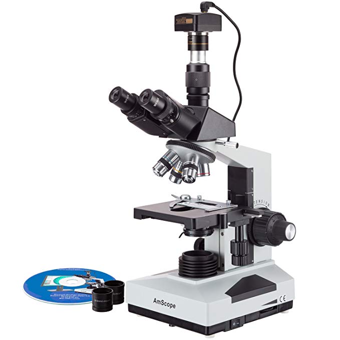
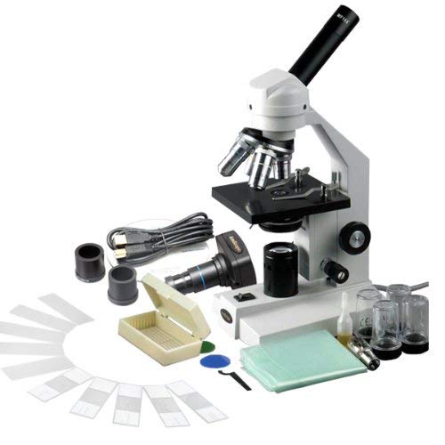
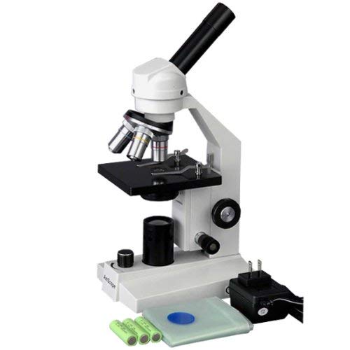
The AmScope M200A-LED cordless monocular compound microscope has interchangeable 10x widefield and 16x widefield eyepieces, a forward-facing nosepiece with three DIN achromatic objectives, LED illumination, coaxial coarse and fine focus, a 0.65 NA single-lens condenser, and a plain stage. The monocular viewing head has a fixed 45-degree vertical inclination to reduce eye and neck strain, and 360-degree rotation capability to enable sharing. The forward-facing revolving nosepiece has 4x, 10x, and 40xS DIN achromatic objectives that provide color correction of magnified images. The 40xS objective is spring loaded to prevent damage to the slide or objective when focusing. A compound microscope is used for inspection and dissection of specimens when two-dimensional images are desired.

The microscope has lower (diascopic) Brightfield illumination that transmits light up through the specimen for enhanced visibility of translucent and transparent objects. Brightfield (BF) illumination allows the specimen to absorb light, resulting in a dark image on a light background. LED illumination provides bright, cool light for working with temperature-sensitive or live specimens, and a rheostat controls the amount of light emanating from the lamp. The 0.65 NA single-lens condenser and disc diaphragm optimize the amount of light illuminating the specimen. The plain stage is 4-3/8 x 4-3/4 inches (W x D; where W is width, the horizontal distance from left to right; D is depth, the horizontal distance from front to back). It has an opening for light transmission, stage clips to secure the slide or specimen in place while viewing, and an upper stage stop to prevent the stage or specimen from coming into contact with the objectives. Separate coaxial coarse and fine focus eases focusing for left- and right-handed viewers, and a rack-and-pinion mechanism provides precise and secure focusing. All the mechanical parts of the microscope are constructed of metal to provide durability and resistance to wear. The cast-alloy metal frame has a stain-resistant enamel finish for durability and to ease cleaning. The microscope can be powered by 110V power supply or three AA batteries (included).

Specifications
Head Monocular
Eyepieces WF10x, WF16x
Lenses 10x, 20x, 40xS DIN achromatic
Stage Plain with clips and stage stop
Focus Coaxial coarse and fine
Condenser Single lens, 0.65 NA
Light source LED with rheostat
Diaphragm Disc
Illumination type Brightfield (BF)
Power 110V or three AA batteries
Overall dimensions 15 x 5-1/8 x 7-1/2 inches (H x W x D)

H is height, the vertical distance from the lowest to highest point; W is width, the horizontal distance from left to right; D is depth, the horizontal distance from front to back.

Microscopes are instruments used to enhance the resolution of an object or image. Types include compound, stereo, or digital. Compound microscopes use a compound optical system with an objective lens and an eyepiece. Stereo microscopes show object depth in a three-dimensional image. Digital microscopes are used to display an image on a monitor, rather than looking through a lens. Microscopes can have monocular (one), binocular (two), or trinocular (three) eyepieces, with varying magnification abilities. Magnification ability refers to the size of an image. Resolution, also known as resolvant power, refers to the clarity of the image. The interaction between field of view (FOV), numerical aperture (NA), and working distance (WD) determines resolution. Microscopes can control magnification through a fixed focus, or through a range of adjustments. They can also utilize LED, fluorescent, and mirror light sources to help control viewing capabilities. Microscopes are widely used in education, lab research, biology, metallurgy, engineering, chemistry, manufacturing, and in the medical, forensic science, and veterinary industries.

United Scope manufactures microscopy equipment and accessories under the brand name AmScope. The company, founded in 1996, is headquartered in Irvine, CA.

What’s in the Box?

  • AmScope M200A compound microscope with plain stage
  • WF10x eyepiece
  • WF16x eyepiece
  • 4x DIN achromatic objective
  • 10x DIN achromatic objective
  • 40xS DIN achromatic objective
  • (2) Stage clips
  • (3) Rechargeable AA batteries
  • Spare tungsten bulbDust cover
  • Instructions

Aven 26700-205 Digital Handheld Microscope, 10x-200x Magnification, Upper UV LED Illumination, With Stand, Includes 1.3MP Camera Review

Aven 26700-205 Digital Handheld Microscope, 10x-200x Magnification, Upper UV LED Illumination, With Stand, Includes 1.3MP Camera

The Aven 26700-205 digital handheld microscope provides 10x to 200x magnification, has a 1.3MP camera, and has a USB 2.0 compatible output for displaying images and video on a computer. Its built-in light source has six UV LEDs operating at 405nm, with a brightness control wheel to illuminate specimens. The microscope has a 1/4″ color CMOS image sensor, and operates on USB power. The included tabletop pole stand allows adjustment of microscope height and magnification. A focus wheel enables optimal resolution. Features include automatic gain control, automatic white balance, and the ability to capture images either using the microscope or a computer (sold separately). A digital handheld lightweight microscope is used in a variety of settings, including classroom and field applications, and where image capture, detailed records, or documentation is required.

The stand is constructed of metal for durability. It is 300 x 200 x 200mm (H x W x D, where H is height, the vertical distance from the lowest to highest point; W is width, the horizontal distance from left to right; and D is depth, the horizontal distance from front to back). The microscope comes with a stand, cable, software, and instructions.

Specifications
Magnification range 10x to 200x
Resolution 1.3MP
Working distance At 10x, 112mm; at 20x, 42mm; at 30x, 19mm; at 40x, 8.5mm
Image sensor 1/4″ CMOS (color)
Image type Still image and video display and capture
Light source Six built-in UV LEDs (405nm)
Microscope stand Metal, table top
Power 5VDC through USB port, 110mA (AVG)
Computer connection USB 2.0
Video format AVI
Cable length 1.8m (6′)
Microscope dimensions 109mm in length and 34.5mm in diameter
Stand dimensions 300 x 200 x 200mm (H x W x D)
Weight 113g

Microscopes are instruments used to enhance the resolution of an object or image. Types include compound, stereo, or digital. Compound microscopes use a compound optical system with an objective lens and an eyepiece. Stereo microscopes show object depth in a three-dimensional image. Digital microscopes are used to display an image on a monitor, rather than looking through a lens. Microscopes can have monocular (one), binocular (two), or trinocular (three) eyepieces, with varying magnification abilities. Magnification ability refers to the size of an image. Resolution, also known as resolvant power, refers to the clarity of the image. The interaction between field of view (FOV), numerical aperture (NA), and working distance (WD) determines resolution. Microscopes can control magnification through a fixed focus, or through a range of adjustments. They can also utilize LED, fluorescent, and mirror light sources to help control viewing capabilities. Microscopes are widely used in education, lab research, biology, metallurgy, engineering, chemistry, manufacturing, and in the medical, forensic science, and veterinary industries.

Aven manufactures optical inspection tools, precision tools, and bench accessories for industrial, scientific, and research applications. The company was founded in 1983 and is headquartered in Ann Arbor, MI.

What’s in the Box?

  • Aven 26700-205 microscope
  • Stand
  • USB cable (attached)
  • CD containing software
  • Instructions

AmScope T490B-3M Digital Compound Trinocular Microscope, WF10x and WF20x Eyepieces, 40X-2000X Magnification, Brightfield, Halogen Illumination, Abbe Condenser, Double-Layer Mechanical Stage, Sliding Head, High-Resolution Optics, Includes 3MP Camera with Reduction Lens and Software Review

AmScope T490B-3M Digital Compound Trinocular Microscope, WF10x and WF20x Eyepieces, 40X-2000X Magnification, Brightfield, Halogen Illumination, Abbe Condenser, Double-Layer Mechanical Stage, Sliding Head, High-Resolution Optics, Includes 3MP Camera with Reduction Lens and Software

    This fits your .
  • Make sure this fits by entering your model number.
  • Digital compound microscope and a 3MP camera with reduction lens and USB 2.0 output for capturing or displaying images on a computer or projector, compatible with Windows XP/Vista/7/8/10 and Mac OSX
  • Trinocular viewing head with vertical camera mount and Siedentopf binocular mount with interchangeable pairs of 10x and 20x widefield eyepieces and sliding head with 55 to 75mm interpupillary adjustment, fixed 30-degree vertical inclination to reduce eye and neck strain, and 360-degree rotation capability to provide a more comprehensive view and enable sharing
  • Forward-facing nosepiece with 4x, 10x, 40xS (spring), and 100xS (spring, oil) DIN achromatic precision-ground optical-glass objectives that provide color correction of magnified images
  • Brightfield, halogen illumination with rheostat to control light intensity, and 1.25 NA Abbe condenser with iris diaphragm and rack-and-pinion focus control for precise focus, clear examination, and light control
  • Double-layer mechanical stage with 1mm stage divisions and stage stop; graduated coarse and fine focus with tension control to prevent stage drift

The AmScope T490B-3M digital compound trinocular microscope has a sliding head, interchangeable pairs of 10x and 20x widefield eyepieces, a forward-facing nosepiece with four DIN achromatic objectives, Brightfield halogen illumination, and a double-layer mechanical stage with a stage stop to protect slides and objectives from damage. The 3MP camera has a CMOS color sensor, a reduction lens, image capture and editing software, and USB 2.0 output to capture or display still or video images on a computer or projector. The trinocular viewing head has a vertical camera mount and a compensation-free binocular viewing head with interchangeable pairs of WF10x and WF20x eyepieces and sliding head to adjust inter-pupillary distance, a fixed 30-degree vertical inclination to reduce eye and neck strain, and a 360-degree rotation capability to provide a more comprehensive view and enable sharing. A Siedentopf binocular head enables the viewer to change the interpupillary distance without changing the tube length, eliminating the need to re-focus the image. It has an interpupillary range of 55 to 75mm to accommodate individual eye differences, and bi-lateral dioptric adjustment accommodates individual eye-strength differences. The vertical trinocular port accepts a camera with a 23mm or C-Mount adapter. The forward-facing nosepiece has 4x, 10x, 40xS (spring), and 100xS (spring, oil) DIN achromatic optical-glass objectives that combine with the eyepieces to provide color correction of magnified images. The 40xS objective is spring loaded to prevent slide damage when focusing. The 100xS spring-loaded oil-immersion objective uses oil between the specimen and the objective lens to provide increased resolution over a standard objective. A digital compound microscope is used for inspection and dissection of specimens when two-dimensional images are desired, and where image capture, detailed records, or documentation is required.

The 3MP digital camera has a CMOS color sensor for displaying still microscopy images and streaming live videos to a computer or projector. The camera has 40x magnification and a 0.5x reduction lens that ensures that the display has a similar field of view as the microscope eyepiece. The camera can be mounted in a C-Mount or any 23mm eye tube. The camera includes image capture and editing software that provides still image and live video capture and editing capability, including measurement functions. The software supports JPG, TIF, GIF, PSD, WMF, and BMP file formats and is compatible with Windows XP, Vista, 7, and 8; Mac OS X; and Linux. Camera drivers are compatible with Windows XP, Vista, 7, and 8; Mac OS X; and Linux. The software includes Windows APIs for native C/C++, C#, DirectShow, Twain, and LabVIEW that enable custom application development. The camera has a USB 2.0 data port (cable included).

The microscope has lower (transmitted, diascopic) Brightfield illumination that transmits light up through the specimen for enhanced visibility of translucent and transparent objects. Brightfield (BF) illumination allows the specimen to absorb light, resulting in a dark image on a light background. Halogen illumination provides bright light in a concentrated path and has a rheostat to control light intensity. The 1.25 NA Abbe condenser is mounted on a rack-and-pinion control system, can be adjusted to control the distance of the light from the stage, and has an iris diaphragm to optimize the amount of light illuminating the specimen. The double-layer mechanical stage, with 1mm stage divisions, locks the slide into place and provides precise slide manipulation along the X- and Y-axis to allow coordinates to be recorded, enabling the viewer to return to a specific location on the slide. A stage stop prevents the stage from coming into contact with the slide and objectives. The stage is 5-1/2 x 5-1/2 inches (140 x 140mm) and has a traveling range of 2-15/16 x 1-3/4 inches (75 x 45mm). Graduated coaxial coarse and fine focus has a focusing range of 1-3/16″ (30mm) and fine-focus divisions of 0.002mm (0.0000787″) that enable measurements to be taken. Focus knob tension control prevents the stage from drifting out of focus. The solid-metal enamel-coated body is durable and resistant to stains and corrosion.

Microscope Specifications
Head Compound trinocular
Magnification 40x-2000x
Trinocular port 23mm or C-Mount
Eyepieces WF10x, WF20x
Objectives 4X, 10x, 40xS, 100xS (oil)
Stage Double-layer mechanical
Lighting configuration Transmitted (lower)
Condenser 1.25 NA Abbe
Diaphragm Iris
Light source Halogen with rheostat, 6V/20W
Illumination type Brightfield
Power 110V

Camera Specifications
Resolution 3MP (2048 x 1536 effective pixels)
Image type Still image and video display and capture
Camera type Brightfield
Camera sensor 1/2″ Aptina MT9T001 CMOS (color)
Magnification 40x
Reduction lens 0.5x
Mounting size 23mm or C-Mount
Frame rate 43fps at 680×510; 22fps at 1024×768; 8fps at 2048×1536
Computer connection USB 2.0 (backward compatible on PCs only)
File formats JPG, TIF, GIF, PSD, WMF, BMP
Software package Image capture and editing for Windows XP, Vista, 7, and 8; Mac OS X; and Linux
Camera driver compatibility Windows XP, Vista, 7, and 8; Mac OS X; and Linux

Microscopes are instruments used to enhance the resolution of an object or image. Types include compound, stereo, or digital. Compound microscopes use a compound optical system with an objective lens and an eyepiece. Stereo microscopes show object depth in a three-dimensional image. Digital microscopes are used to display an image on a monitor, rather than looking through a lens. Microscopes can have monocular (one), binocular (two), or trinocular (three) eyepieces, with varying magnification abilities. Magnification ability refers to the size of an image. Resolution, also known as resolvant power, refers to the clarity of the image. The interaction between field of view (FOV), numerical aperture (NA), and working distance (WD) determines resolution. Microscopes can control magnification through a fixed focus, or through a range of adjustments. They can also utilize LED, fluorescent, and mirror light sources to help control viewing capabilities. Microscopes are widely used in education, lab research, biology, metallurgy, engineering, chemistry, manufacturing, and in the medical, forensic science, and veterinary industries.

United Scope manufactures microscopy equipment and accessories under the brand name AmScope. The company, founded in 1996, is headquartered in Irvine, CA.

What’s in the Box?

  • AmScope T490B-3M microscope with double-layer mechanical stage
  • WF10x eyepieces, 23mm, one pair
  • WF20x eyepieces, 23mm, one pair
  • 4x DIN achromatic objective
  • 10x DIN achromatic objective, 20mm
  • 40xS DIN achromatic objective, 20mm
  • 100xS (oil) DIN achromatic objective, 20mm
  • Immersion oil, one bottle
  • (3) Color filters; blue, green, and yellow
  • 3MP digital camera (MU300)
  • 0.5x reduction lens
  • USB 2.0 cable
  • Software CD
  • Spare halogen bulb
  • Spare fuse
  • Dust cover
  • Power cord
  • Instructions

7X-45X Dual Lit 6W LED Trinocular Stereo Zoom Microscope Review

7X-45X Dual Lit 6W LED Trinocular Stereo Zoom Microscope

    This fits your .
  • Make sure this fits by entering your model number.
  • Super widefield optical system offers crisp clear stereo images
  • Both top and bottom LED illuminations
  • 7X-45X zoom magnification power
  • Large working distance; large field of view; sturdy metal tracking stand
  • Trinocular port for adding photo or video capability

This top of the line 7X-45X trinocular stereo zoom microscope is equipped with SUPER widefield optics, and a versatile dual LED illumination system. This microscope has been designed for use in organic or temperature-sensitive applications – like, dissecting live tissues or observing live organisms. Our low-temperature LED system allows you to use both incident illumination, which shines light down onto a 3-dimensional specimen, and reflecting-plate bottom illumination, which shines light up through a transparent sample. Because the top and bottom LED lights are controlled by separate dimmers, you are able to customize lighting for the optimal level of illumination, whatever the situation.

The optics of this SM series microscope features a large zoom magnification range, which produces sharp, crystal-clear stereo images, while providing a generous working distance. The 360 degree swiveling, trinocular head is attached at a 45 degree angle, which ensures easy and comfortable observation – especially when used in conjunction with the included rubber eyeguards. On the base, the LED reflecting-plate measures 3-3/4″ or 95 mm in diameter.

This microscope is especially useful for applications that require cool illumination. It is perfect for the examination of organic matter, such as insects, plants, or virtually any 3-dimensional, temperature-sensitive object. It is ideal for those who work in biology, forestry, archaeology, and agriculture. With its high resolution, good depth, and broad field of view, this microscope can be used for both educational and professional purposes in research labs, hospitals, schools, and veterinary offices.

This microscope provides all the excellent features of a high quality laboratory instrument at an affordable price. It comes brand new in a factory-sealed box.

AmScope SM-3BZZ-80S Professional Binocular Stereo Zoom Microscope, WH10x and WH20x Eyepieces, 3.5X-180X Magnification, 0.7X-4.5X Zoom Objective, 80-Bulb LED Ring Light, Single-Arm Boom Stand, 90V-265V, Includes 0.5x and 2.0x Barlow Lens Review

AmScope SM-3BZZ-80S Professional Binocular Stereo Zoom Microscope, WH10x and WH20x Eyepieces, 3.5X-180X Magnification, 0.7X-4.5X Zoom Objective, 80-Bulb LED Ring Light, Single-Arm Boom Stand, 90V-265V, Includes 0.5x and 2.0x Barlow Lens

    This fits your .
  • Make sure this fits by entering your model number.
  • Professional stereo microscope with boom stand has long working distance to enable users to perform work or manipulate large items, including circuit boards and dental appliances
  • Binocular viewing head with interchangeable pairs of 10x and 20x super-widefield eyepieces, adjustable interpupillary distance, fixed 45-degree vertical inclination to reduce eye and neck strain, and 360-degree rotation capability to enable sharing
  • 0.7x-4.5x zoom objective provides continuous zoom magnification and longer focal length for inspecting large-scale specimens, a 05.x Barlow lens extends the working distance, and a 2.0x Barlow lens extends the magnification range
  • Removable rheostat-controlled 80-bulb LED ring light provides intensity control and bright, cool light for working with temperature-sensitive or live specimens
  • Single boom arm with 8″ vertical working distance and adjustable 16″ boom arm enables users to adjust the microscope on the X- and Y-axes

The AmScope SM-3BZZ-80S professional stereo zoom microscope has interchangeable pairs of 10x and 20x super-widefield high-eyepoint eyepieces, a 0.7x-4.5x zoom objective, two Barlow lenses, and a single-arm boom stand. The microscope has an overall magnification range of 3.5x-180x. The binocular viewing head has an interpupillary range of 55 to 75mm, a 45-degree inclination to reduce eye and neck strain, and 360-degree rotation to enable sharing. The WH10x20mm and WH20x10mm super-widefield high-eyepoint eyepieces combine with the 0.7x-4.5x zoom objective to provide continuous zoom magnification and a longer working distance for inspecting large-scale specimens that require handling or repair. High-eyepoint eyepieces ease viewing for users who wear glasses, and dioptric adjustment accommodates individual eye-strength differences. The microscope includes 0.5x and 2.0x Barlow lenses that can be added to the objective to increase the magnification range. A Barlow lens with a magnification of less than 1.0 reduces magnification and increases the working distance, while a Barlow lens with a magnification greater than 1.0 increases the magnification and reduces the working distance. A stereo microscope, sometimes called an inspection or dissection microscope, has low magnification and a long working distance that enables users to manipulate the object being inspected.

The removable 80-bulb LED ring light provides bright, cool light for working with temperature-sensitive or live specimens. The ring light provides shadow-free illumination, and a rheostat controls the amount of light emanating from the lamp. Power and adjustment controls are located on the light head. The light has a 100,000-hour life span and a 90V-265V variable power supply. The microscope has a 17″ pillar that has an 8″ vertical working distance. The 16″ single-arm boom has a 24″ overall length. Bilateral focus eases use for left- and right-handed users. The microscope head can be rotated on the X- and Y-axes. The solid cast-steel base is 1-3/4 x 9 x 9 inches (H x W x D, where H is height, the vertical distance from the lowest to highest point; W is width, the horizontal distance from left to right; D is depth, the horizontal distance from front to back).

Specifications
Head Binocular
Magnification range 3.5x-180x
Zoom objective power 0.7x-4.5x
Eyepieces (DIN, 30mm) WH10x20mm and WH20x10mm high-eyepoint
Zoom range 52:1
Field of view 2-1/2″
Optical working distance Up to 8″
Microscope stand Single-arm boom stand
Head movement X- and Y-axes
Illumination type Episcopic (reflected)
Light source 80-bulb LED ring light
Power 90V-265V

Microscopes are instruments used to enhance the resolution of an object or image. Types include compound, stereo, or digital. Compound microscopes use a compound optical system with an objective lens and an eyepiece. Stereo microscopes show object depth in a three-dimensional image. Digital microscopes are used to display an image on a monitor, rather than looking through a lens. Microscopes can have monocular (one), binocular (two), or trinocular (three) eyepieces, with varying magnification abilities. Magnification ability refers to the size of an image. Resolution, also known as resolvant power, refers to the clarity of the image. The interaction between field of view (FOV), numerical aperture (NA), and working distance (WD) determines resolution. Microscopes can control magnification through a fixed focus, or through a range of adjustments. They can also utilize LED, fluorescent, and mirror light sources to help control viewing capabilities. Microscopes are widely used in education, lab research, biology, metallurgy, engineering, chemistry, manufacturing, and in the medical, forensic science, and veterinary industries.

United Scope manufactures microscopy equipment and accessories under the brand name AmScope. The company, founded in 1996, is headquartered in Irvine, CA.

What’s in the Box?

  • AmScope SM-3BZZ-80S stereo zoom microscope with single-arm boom stand
  • WH10x20mm eyepieces, 30mm, one pair
  • WH20x10mm eyepieces, 30mm, one pair
  • 0.7x-4.5x zoom objective
  • 0.5x Barlow lens
  • 2.0x Barlow lens
  • 80-bulb LED ring light (LED-80S)
  • Focus rack
  • Eye guards, one pair
  • Instructions

AmScope SM-3TZ-54S-5M Digital Professional Trinocular Stereo Zoom Microscope, WH10x Eyepieces, 3.5X-90X Magnification, 0.7X-4.5X Zoom Objective, 54-Bulb LED Light, Single-Arm Boom Stand, 110V-240V, Includes 0.5X and 2.0X Barlow Lenses and 5MP Camera with Reduction Lens and Software Review

AmScope SM-3TZ-54S-5M Digital Professional Trinocular Stereo Zoom Microscope, WH10x Eyepieces, 3.5X-90X Magnification, 0.7X-4.5X Zoom Objective, 54-Bulb LED Light, Single-Arm Boom Stand, 110V-240V, Includes 0.5X and 2.0X Barlow Lenses and 5MP Camera with Reduction Lens and Software

    This fits your .
  • Make sure this fits by entering your model number.
  • Digital professional stereo microscope with boom stand has long working distance to enable users to perform work or manipulate large items, including circuit boards and dental appliances. Includes 5MP camera, reduction lens, USB 2.0 and software capturing or displaying images. Compatible with Windows 10/XP/Vista/7/8/10 and Mac OS X.
  • Trinocular viewing head with pair of 10x super-widefield eyepieces, adjustable interpupillary distance, fixed 45-degree vertical inclination to reduce eye and neck strain, and 360-degree rotation capability to enable sharing
  • 0.7x-4.5x zoom objective provides continuous zoom magnification and longer focal length for inspecting large-scale specimens, a 0.5x Barlow lens extends the working distance, and a 2.0x Barlow lens extends the magnification range
  • Removable rheostat-controlled 54-bulb LED ring light provides intensity control and bright, cool light for working with temperature-sensitive or live specimens
  • Single boom arm with 8″ vertical working distance and adjustable 16″ boom arm enables users to adjust the microscope on the X- and Y-axes

The AmScope SM-3TZ-54S-5M digital professional stereo zoom microscope has a pair of 10x super-widefield high-eyepoint eyepieces, a 0.7x-4.5x zoom objective, and a single-arm boom stand. The microscope has an overall magnification range of 3.5x-90x. The 5MP camera has a CMOS color sensor, a reduction lens, image capture and editing software, and USB 2.0 output to capture or display still or video images on a computer or projector. The trinocular viewing head has an interpupillary range of 55 to 75mm, a 45-degree inclination to reduce eye and neck strain, and 360-degree rotation to enable sharing. The vertical trinocular port can be used as a C-Mount or 23mm photo port. The WH10x20mm super-widefield high-eyepoint eyepieces combine with the 0.7x-4.5x zoom objective to provide continuous zoom magnification and a longer working distance for inspecting large-scale specimens that require handling or repair. The microscope comes with 0.5x and 2.0x Barlow lenses that can be added to the objective to increase the magnification range. A Barlow lens with a magnification of less than 1.0 reduces magnification and increases the working distance, while a Barlow lens with a magnification greater than 1.0 increases the magnification and reduces the working distance. High-eyepoint eyepieces ease viewing for users who wear glasses, and dioptric adjustment accommodates individual eye-strength differences. A digital stereo microscope, sometimes called an inspection or dissection microscope, has low magnification and a long working distance that enables users to manipulate the object being inspected, and is used where image capture, detailed records, or documentation is required.

The 5MP digital camera has a CMOS color sensor for displaying still microscopy images and streaming live videos to a computer or projector. The camera has 40x magnification and a 0.5x reduction lens that ensures that the display has a similar field of view as the microscope eyepiece. The camera can be mounted in a C-Mount or any 23mm eye tube. The camera includes image capture and editing software that provides still image and live video capture and editing capability, including measurement functions. The software supports JPG, TIF, GIF, PSD, WMF, and BMP file formats and is compatible with Windows 10/XP/Vista/7/8/10 and Mac OS X. Camera drivers are compatible withWindows 10/XP/Vista/7/8/10 and Mac OS X. The software includes Windows APIs for native C/C++, C#, DirectShow, Twain, and LabVIEW that enable custom application development. The camera has a USB 2.0 data port (cable included).

The removable 54-bulb LED ring light provides bright, cool light for working with temperature-sensitive or live specimens. The ring light provides shadow-free illumination, and a rheostat controls the amount of light emanating from the lamp. Power and adjustment controls are located on the light head. The light uses a 110V-240V variable power supply. The microscope has a 17″ pillar that has an 8″ vertical working distance. The 16″ single-arm boom has a 24″ overall length. Bilateral focus eases use for left- and right-handed users. The microscope head can be rotated on the X- and Y-axes. The solid cast-steel base is 1-3/4 x 9 x 9 inches (H x W x D, where H is height, the vertical distance from the lowest to highest point; W is width, the horizontal distance from left to right; D is depth, the horizontal distance from front to back).

Microscope Specifications
Head Trinocular
Magnification range 3.5x-90x
Zoom objective power 0.7x-4.5x
Eyepieces (DIN, 30mm) WH10x20mm high-eyepoint
Trinocular port C-Mount or 23mm
Field of view 2-1/2″
Optical working distance Up to 8″
Microscope stand Single-arm boom stand
Head movement X- and Y-axes
Illumination type Episcopic (reflected)
Light source 54-bulb LED ring light
Power 110V-240V

Camera Specifications
Resolution 5MP (2592 x 1944 effective pixels)
Image type Still image and video display and capture
Camera type Brightfield
Camera sensor 1/2.5″ Aptina MT9P001 CMOS (color)
Magnification 40x
Reduction lens 0.5x
Mounting size 23mm or C-Mount
Frame rate 60fps at 640×480; 18fps at 1280×960; 5fps at 2592×1944
Computer connection USB 2.0 (backward compatible on PCs only)
File formats JPG, TIF, GIF, PSD, WMF, BMP
Software package Image capture and editing for Windows XP, Vista, 7, and 8; Mac OS X; and Linux
Camera driver compatibility Windows XP, Vista, 7, and 8; Mac OS X; and Linux

Microscopes are instruments used to enhance the resolution of an object or image. Types include compound, stereo, or digital. Compound microscopes use a compound optical system with an objective lens and an eyepiece. Stereo microscopes show object depth in a three-dimensional image. Digital microscopes are used to display an image on a monitor, rather than looking through a lens. Microscopes can have monocular (one), binocular (two), or trinocular (three) eyepieces, with varying magnification abilities. Magnification ability refers to the size of an image. Resolution, also known as resolvant power, refers to the clarity of the image. The interaction between field of view (FOV), numerical aperture (NA), and working distance (WD) determines resolution. Microscopes can control magnification through a fixed focus, or through a range of adjustments. They can also utilize LED, fluorescent, and mirror light sources to help control viewing capabilities. Microscopes are widely used in education, lab research, biology, metallurgy, engineering, chemistry, manufacturing, and in the medical, forensic science, and veterinary industries.

United Scope manufactures microscopy equipment and accessories under the brand name AmScope. The company, founded in 1996, is headquartered in Irvine, CA.

What’s in the Box?

  • AmScope SM-3TZ-54S-5M stereo zoom microscope with single-arm boom stand
  • WH10x20mm eyepieces, 30mm, one pair
  • 0.7x-4.5x zoom objective
  • 0.5x Barlow lens
  • 2.0x Barlow lens
  • 54-Bulb LED ring light (LED-54S)
  • Focus rack
  • 5MP digital camera (MU500)
  • 0.5x reduction lens
  • USB 2.0 cable
  • Software CD
  • Eye guards, one pair
  • Instructions

AmScope SE305R-PX-LED Forward-Mounted Binocular Stereo Microscope, WF5x and WF10x Eyepieces, 5X/10X/15X/30X Magnification, 1X and 3X Objectives, Upper and Lower LED Lighting, Reversible Black/White Stage Plate, Pillar Stand, 120V or Battery-Powered Review

AmScope SE305R-PX-LED Forward-Mounted Binocular Stereo Microscope, WF5x and WF10x Eyepieces, 5X/10X/15X/30X Magnification, 1X and 3X Objectives, Upper and Lower LED Lighting, Reversible Black/White Stage Plate, Pillar Stand, 120V or Battery-Powered

    This fits your .
  • Make sure this fits by entering your model number.
  • Stereo microscope for home or classroom use enables students and hobbyists to inspect biological specimens, rocks, stamps, jewelry, and large specimens that require handing or manipulation
  • Forward-mounted binocular viewing head with interchangeable pairs of 5x and 10x widefield eyepieces, adjustable interpupillary distance, and 45-degree inclination to simplify viewing for young users
  • Interchangeable 1x and 3x objectives provide low magnification and longer focal length for inspecting large-scale specimens
  • Upper and lower illumination has LED light source to illuminate specimens
  • Frosted plate allows light to pass through from below with transparent specimens, reversible black and white stage plate provides contrast with light- and dark-colored specimens, and stage clips secure the specimen during viewing

The AmScope SE305R-PX-LED stereo microscope has interchangeable pairs of 5x18mm and 10x18mm widefield eyepieces, 1x and 3x objectives, upper and lower LED lighting, two stage plates, and a pillar stand. The forward-mounted binocular viewing head has an interpupillary range of 55 to 75mm and a 45-degree inclination to ease viewing for young users. The WF5x18mm and WF10x18mm eyepieces combine with the 1x and 3x objectives to provide 5x, 10x, 15x, and 30x magnification, and a longer working distance for inspecting large-scale specimens that require handling or repair. The optical glass lenses provide sharp images, and are fully coated to ensure high-resolution images. Dioptric adjustment accommodates individual eye-strength differences, and eye guards ensure comfortable viewing. A stereo microscope, sometimes called an inspection or dissection microscope, has low magnification and a long working distance that enables users to manipulate the object being inspected.

The microscope has upper (episcopic) illumination that reflects light off the specimen for enhanced visibility of opaque specimens, and lower (diascopic) illumination that transmits light up through the specimen for enhanced visibility of translucent and transparent objects. The LED light source provides bright, cool light for working with temperature-sensitive or live specimens. The pillar-style stand secures the head for precise focusing, and bilateral coarse focus eases use for left- and right-handed users. The stage has a frosted stage plate for use with lower (transmitted) lighting, and a reversible black and white stage plate that provides contrast with light- and dark-colored specimens when used with upper (reflected) lighting. Stage clips that secure the specimen during viewing. The microscope has a 2-1/8″ (53mm) working distance. All metal construction is durable and stain-resistant.

Specifications
Head Forward-mounted binocular
Magnification 5x, 10x, 15x, 30x
Objective power 1x, 3x
Eyepieces (DIN, 30.5mm) WF5x18mm,
WF10x18mm
Working distance 2-1/8″ (53mm)
Focus Bilateral coarse
Microscope stand Pillar stand
Stage plate (95mm) Frosted and reversible black/white
Illumination type Upper (reflected) and lower (transmitted)
Light source LED
Power 120V, UL listed

Microscopes are instruments used to enhance the resolution of an object or image. Types include compound, stereo, or digital. Compound microscopes use a compound optical system with an objective lens and an eyepiece. Stereo microscopes show object depth in a three-dimensional image. Digital microscopes are used to display an image on a monitor, rather than looking through a lens. Microscopes can have monocular (one), binocular (two), or trinocular (three) eyepieces, with varying magnification abilities. Magnification ability refers to the size of an image. Resolution, also known as resolvant power, refers to the clarity of the image. The interaction between field of view (FOV), numerical aperture (NA), and working distance (WD) determines resolution. Microscopes can control magnification through a fixed focus, or through a range of adjustments. They can also utilize LED, fluorescent, and mirror light sources to help control viewing capabilities. Microscopes are widely used in education, lab research, biology, metallurgy, engineering, chemistry, manufacturing, and in the medical, forensic science, and veterinary industries.

United Scope manufactures microscopy equipment and accessories under the brand name AmScope. The company, founded in 1996, is headquartered in Irvine, CA.

What’s in the Box?

  • AmScope SE305R-PX-LED stereo microscope with forward head and pillar stand
  • WF5x18mm eyepieces, 30.5mm, one pair
  • WF10x18mm eyepieces, 30.5mm, one pair
  • 1x objective
  • 3x objective
  • Black/white stage plate, 95mm
  • Frosted stage plate, 95mm
  • (2) Eye guards
  • (2) Stage clips
  • LED bulb
  • Focusing wrench
  • Dust cover
  • Power cord
  • Instructions

AmScope M500A-P-PB10 Digital Monocular Compound Microscope, WF10x and WF16x Eyepieces, 40x-1600x Magnification, Anti-Mold Optics, Tungsten Illumination, Brightfield, Abbe Condenser, Coarse and Fine Focus, Plain Stage, 110V, Includes 0.3MP Camera with Reduction Lens and Software, 5 Blank Slides, and 5 Prepared Slides Review

AmScope M500A-P-PB10 Digital Monocular Compound Microscope, WF10x and WF16x Eyepieces, 40x-1600x Magnification, Anti-Mold Optics, Tungsten Illumination, Brightfield, Abbe Condenser, Coarse and Fine Focus, Plain Stage, 110V, Includes 0.3MP Camera with Reduction Lens and Software, 5 Blank Slides, and 5 Prepared Slides

    This fits your .
  • Make sure this fits by entering your model number.
  • Digital compound microscope and a 0.3MP camera with reduction lens and USB 2.0 output for capturing or displaying images on a computer or projector
  • Monocular viewing head with interchangeable 10x and 16x widefield eyepieces, fixed 45-degree vertical inclination to reduce eye and neck strain, 360-degree rotation capability to enable sharing, and anti-mold head to preserves optics in high-humidity area
  • Forward-facing nosepiece with precision-ground 4x, 10x, 40xS, and 100xS (oil) DIN achromatic glass objectives provide color correction of magnified images
  • Brightfield imaging with rheostat-controlled tungsten (incandescent) illumination and spiral-control 1.25 NA Abbe condenser with iris diaphragm and color filter
  • Plain stage with clips to secure specimen, coaxial coarse and fine focus on rack-and-pinion mechanism to provide precise focus, and tension control to prevent stage drift

The AmScope M500A-P-PB10 digital monocular compound microscope has interchangeable 10x and 16x widefield eyepieces, a forward-facing nosepiece with four DIN achromatic objectives, tungsten illumination, coaxial separate coarse and fine focus, a 1.25 NA Abbe condenser, and a plain stage. The 0.3MP camera has a CMOS color sensor, a reduction lens, image capture and editing software, and USB 2.0 output to capture or display still or video images on a computer or projector. The monocular viewing head has a fixed 45-degree vertical inclination to reduce eye and neck strain, and 360-degree rotation capability to enable sharing. An anti-mold head preserves optics in high-humidity areas. The forward-facing revolving nosepiece has 4x, 10x, 40xS, and 100xS (oil) DIN achromatic objectives that provide color correction of magnified images. The 40xS and 100xS objectives are spring loaded to prevent damage to the slide or objective when focusing. The 100xS oil-immersion objective uses oil between the specimen and the objective lens to provide increased resolution over a standard objective. The fully-coated optical system provides sharp, high-resolution images. A digital compound microscope is used for inspection and dissection of specimens when two-dimensional images are desired, and where image capture, detailed records, or documentation is required.

The 0.3MP digital camera has a CMOS color sensor for displaying still microscopy images and streaming live videos to a computer or projector. The camera has 40x magnification and a 0.37x reduction lens that ensures that the display has a similar field of view as the microscope eyepiece. The camera can be mounted in a C-Mount or any 23mm eye tube. The camera includes image capture and editing software that provides still image and live video capture and editing capability, including measurement functions. The software supports JPG, TIF, GIF, PSD, WMF, and BMP file formats and is compatible with Windows XP, Vista, 7, and 8; Mac OS X; and Linux. Camera drivers are compatible with Windows XP, Vista, 7, and 8; Mac OS X; and Linux. The software includes Windows APIs for native C/C++, C#, DirectShow, Twain, and LabVIEW that enable custom application development. The camera has a USB 2.0 data port (cable included).

The microscope has lower (diascopic) Brightfield illumination that transmits light up through the specimen for enhanced visibility of translucent and transparent objects. Brightfield (BF) illumination allows the specimen to absorb light, resulting in a dark image on a light background. Tungsten (incandescent) illumination provides bright light, and a rheostat controls the amount of light emanating from the lamp. The 1.25 NA Abbe condenser can be adjusted to control the distance of the light from the stage and has an iris diaphragm to optimize the amount of light illuminating the specimen. The condenser is controlled using a spiral mechanism. The plain stage has clips to secure the specimen in place, and is 4-3/8 x 4-3/4 inches (110 x 120mm) (W x D; where W is width, the horizontal distance from left to right; D is depth, the horizontal distance from front to back). Separate coaxial coarse and fine focus eases focusing for left- and right-handed viewers, and a rack-and-pinion mechanism provides precise and secure focusing. Focus tension control prevents stage drift. All the mechanical parts of the microscope are constructed of metal to provide durability and resistance to wear. The metal frame has a stain-resistant enamel finish for durability and to ease cleaning. The microscope includes five blank and five prepared slides.

Microscope Specifications
Head Monocular
Eyepieces WF10x, WF16x (23mm)
Lenses 10x, 20x, 40xS, 100xS (oil) DIN achromatic (20mm)
Stage Plain stage with stage clips
Focus Coaxial coarse and fine
Condenser 1.25 NA Abbe
Light source Tungsten with rheostat, 20W
Diaphragm Iris
Illumination type Brightfield (BF)
Power 110V

Camera Specifications
Resolution 0.3MP (640 x 480 effective pixels)
Image type Still image and video display and capture
Camera type Brightfield
Camera sensor 1/4″ Aptina CMOS (color)
Magnification 40x
Reduction lens 0.37x
Mounting size 23mm or C-Mount
Frame rate 79fps at 320×240; 30fps at 640×480
Computer connection USB 2.0
File formats JPG, TIF, GIF, PSD, WMF, BMP
Software package Image capture and editing for Windows XP, Vista, 7, and 8; Mac OS X; and Linux
Camera driver compatibility Windows XP, Vista, 7, and 8; Mac OS X; and Linux

Microscopes are instruments used to enhance the resolution of an object or image. Types include compound, stereo, or digital. Compound microscopes use a compound optical system with an objective lens and an eyepiece. Stereo microscopes show object depth in a three-dimensional image. Digital microscopes are used to display an image on a monitor, rather than looking through a lens. Microscopes can have monocular (one), binocular (two), or trinocular (three) eyepieces, with varying magnification abilities. Magnification ability refers to the size of an image. Resolution, also known as resolvant power, refers to the clarity of the image. The interaction between field of view (FOV), numerical aperture (NA), and working distance (WD) determines resolution. Microscopes can control magnification through a fixed focus, or through a range of adjustments. They can also utilize LED, fluorescent, and mirror light sources to help control viewing capabilities. Microscopes are widely used in education, lab research, biology, metallurgy, engineering, chemistry, manufacturing, and in the medical, forensic science, and veterinary industries.

United Scope manufactures microscopy equipment and accessories under the brand name AmScope. The company, founded in 1996, is headquartered in Irvine, CA.

What’s in the Box?

  • AmScope M500A-P-PB10 microscope with plain stage
  • WF10x eyepiece, 23mm
  • WF16x eyepiece, 23mm
  • 4x DIN achromatic objective, 20mm
  • 10x DIN achromatic objective, 20mm
  • 40xS DIN achromatic objective, 20mm
  • 100xS (oil) DIN achromatic objective, 20mm
  • (2) Stage clips
  • 0.3MP digital camera (MU035)
  • 0.37x reduction lens
  • Software CD
  • USB 2.0 cable
  • (5) Blank slides
  • (5) Prepared slides
  • Wrench
  • Color filter
  • Immersion oil
  • Dust cover
  • Instructions

AIHOME 14MP HD TV HDMI USB Industry Digital C-mount Microscope Camera TF Card + 180x Zoom C-MOUNT Lens Review

AIHOME 14MP HD TV HDMI USB Industry Digital C-mount Microscope Camera TF Card + 180x Zoom C-MOUNT Lens

    This fits your .
  • Make sure this fits by entering your model number.
  • 14 megapixel Panasonic sensor 1/2.3 inch
  • It build in digital crosshairs and color control, add line in the pictures
  • Transverse and vertical line: Support multi-color, 5 pcs of transverse lines/vertical lines, movable
  • Image resolution: 4320×3240 (for TF card) 1920×1080 @ 60FPS(for TF card);1920×1080 @ 30FPS(for USB)
  • Video format: MP4(for TF card) ; Image format: JPG; USB Video resolution: 1920×1080 @ 30FPS

Features:

Image sensor: 14 megapixel Panasonic sensor 1/2.3 inch
Effective pixel: 14 megapixel
Pixel size: 1.43*1.43um
Frame rate: 60fps
Definition: FULL HD
Housing: metal
Lens:C S for test, custom provide
White balance: Auto/manual
Digital magnification: 5 times digital zoom
Brightness control: Auto/manual
Color: R/G/B adjustable
Freeze: Support
OSD: Support
Film and record: Support Cross cursor”Support multi-color, size adjustable
Transverse and vertical line:Support multi-color, 4 pcs of transverse lines/vertical lines, movable
SNR: Support
TF card interface: Max 64G
HDMI interface: Standard HDMI output (Type A)
Lens: C
USB interface : Standard usb2.0 interface (Type B)

Specification:

Model: Camera with HDMI USB2.0 two output
Image format: jpg
Image resolution: 4320×3240(14MP) (for TF card)
Video format: MP4
USB Video resolution: 1920×1080 @ 30FPS
White Balance: Auto
Light: Auto
Color: Color
Negative: Support
Morror: Left/right,Up/Down
Freeze: Support
OSD: English/Chinese/French/Spanish/Portuguese
Line: different color,4 horizontal line , 4 vertical line,any positions

C-MOUNT Lens:

Working distance: 100mm-150mm
Zoom C-mount Lens
0.5X C-mount adapter
zoom ratios: 6.5:1
Objective Magnification Power by 0.7 – 4.5X(about 10 – 180X on the display)
Size: 168mm(L) * 50mm(DIA)
Weight: 450g

Package Including:

1 x Microscope Camera
1 x Power supply
1 x USB line
1 x Software download
1 x 180x Lens

Summit SK2-14X – 14.0MP – Digital USB 2.0 Microscope Camera – PC/MAC Compatible – Image Capture Software – Measuring Software (PC only) – C-Mount – 23mm Eyepiece Adapter Review

Summit SK2-14X - 14.0MP - Digital USB 2.0 Microscope Camera - PC/MAC Compatible - Image Capture Software - Measuring Software (PC only) - C-Mount - 23mm Eyepiece Adapter

The new OptixCam Summit K2 Series 14.0MP digital microscope camera is specifically designed as a cost-effective and versatile solution for the wide-ranging imaging requirements of life science researchers, healthcare professionals, industrial inspectors and other disciplines. 14.0MP resolution offers excellent details for images projected on large screens or when printing with large-format applications. Windows PC and MAC users benefit from easy-to-use feature-rich software, accurate color, clarity and image detail, while low noise USB 2.0 data transfer maximizes workflow and productivity.

All Summit K2 microscope cameras share a rugged aluminum alloy body fitted with different size Aptina CMOS sensors depending on the model. Advanced documentation and measuring software is included along with a C-mount adapter with 23mm smooth bore and 0.5X reducing lens to more closely match the captured image with the eyepiece view.

The included K2View software for Windows creates a complete imaging solution with intuitive camera controls, image capture and advanced options like annotation, geometric measuring and storage in JPG, BMP, TIFF, PNG, PCX AND TARGA format. The software also includes auto white balance, color enhancement, anti-flicker and other benefits. TWAIN and DirectX plug-ins are also included on the installation CD and are independent of the main driver, allowing discreet control of the camera for these applications.

Scroll To Top