AmScope ME300TZB-2L-8M Digital Episcopic and Diascopic Trinocular Metallurgical Microscope, WF10x and WF20x Eyepieces, 40X-2000X Magnification, Halogen Illumination with Rheostat, Double-Layer Mechanical Stage, Sliding Head, High-Resolution Optics, Includes 8MP Camera with Reduction Lens and Software Review

AmScope ME300TZB-2L-8M Digital Episcopic and Diascopic Trinocular Metallurgical Microscope, WF10x and WF20x Eyepieces, 40X-2000X Magnification, Halogen Illumination with Rheostat, Double-Layer Mechanical Stage, Sliding Head, High-Resolution Optics, Includes 8MP Camera with Reduction Lens and Software

    This fits your .
  • Make sure this fits by entering your model number.
  • Digital metallurgical microscope uses transmitted and reflected light to view metallic specimens, including electronics, or larger specimens not viewable on a standard microscope stage, and an 8MP camera with reduction lens and USB 2.0 output for capturing or displaying images on a computer or projector
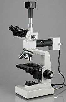
  • Trinocular head with interchangeable pairs of 10x and 20x widefield eyepieces and sliding head with 55 to 75mm interpupillary adjustment, fixed 30-degree vertical inclination to reduce eye and neck strain, and 360-degree rotation capability to enable sharing
  • Forward-facing quadruple nosepiece with 4x, 10x, 40xS, and 100xS (oil) DIN plan optical-glass objectives that provide improved focus over the entire field of view
  • Episcopic (reflected) and diascopic (transmitted) illumination system has halogen illumination with rheostat to control light intensity
  • Double-layer mechanical stage has 1mm stage divisions with 0.1mm vernier graduations and stage stop; graduated coarse and fine focus with tension control to prevent stage drift

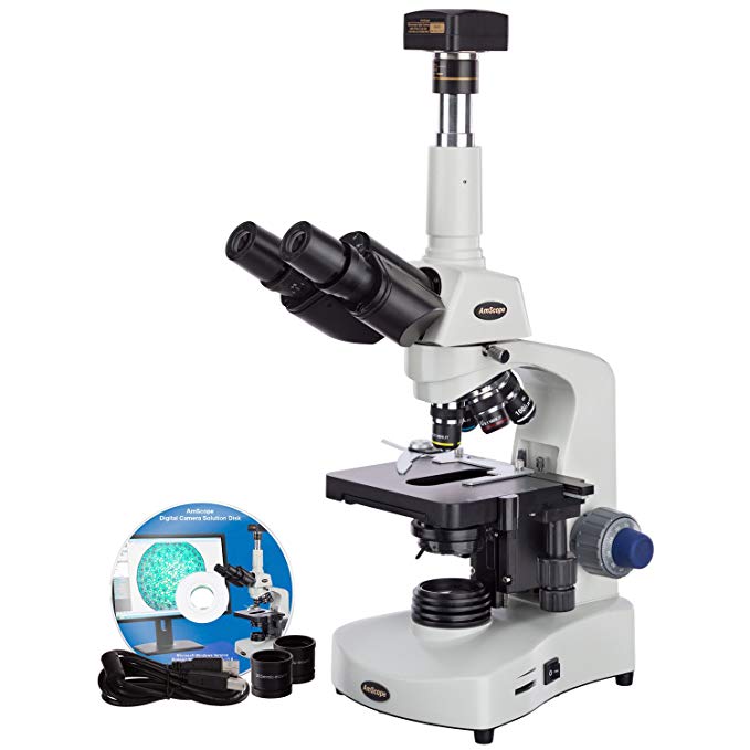
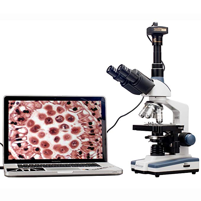
The AmScope ME300TZB-2L-8M digital metallurgical trinocular microscope has interchangeable pairs of 10x and 20x widefield eyepieces, a forward-facing quadruple nosepiece with four DIN achromatic objectives, Brightfield episcopic and diascopic halogen illumination, and a double-layer mechanical stage with a stage stop to protect slides and objectives from damage. The 8MP camera has a CMOS color sensor, a reduction lens, image capture and editing software, and USB 2.0 output to capture or display still or video images on a computer or projector. The trinocular viewing head has a 23mm vertical camera mount, a sliding binocular head to adjust interpupillary distance, a fixed 30-degree vertical inclination to reduce eye and neck strain, and a 360-degree rotation capability to enable sharing. The sliding head has a range of 55 to 75mm to accommodate individual eye differences, and bilateral dioptric adjustment to accommodate individual eye-strength differences. The forward-facing nosepiece has 4x, 10x, 40xS (spring), and 100xS (oil) DIN plan optical-glass objectives that combine with the eyepieces to provide color correction of magnified images. The 40xS objective is spring loaded to prevent slide damage when focusing. The 100xS spring-loaded oil-immersion objective uses oil between the specimen and the objective lens to provide increased resolution over a standard objective. Plan objectives provide improved focus over the entire range of the viewing field. A digital metallurgical microscope uses transmitted and reflected light to view opaque or metallurgical specimens, or larger specimens that cannot be viewed on a standard microscope stage, and is used where image capture, detailed records, or documentation is required.

The 8MP digital camera has a CMOS color sensor for displaying still microscopy images and streaming live videos to a computer or projector. The camera has 40x magnification and a 0.5x reduction lens that ensures that the display has a similar field of view as the microscope eyepiece. The camera can be mounted in a C-Mount or any 23mm eye tube. The camera includes image capture and editing software that provides still image and live video capture and editing capability, including measurement functions. The software supports JPG, TIF, GIF, PSD, WMF, and BMP file formats and is compatible with Windows XP, Vista, 7, and 8; Mac OS X; and Linux. Camera drivers are compatible with Windows XP, Vista, 7, and 8; Mac OS X; and Linux. The software includes Windows APIs for native C/C++, C#, DirectShow, Twain, and LabVIEW that enable custom application development. The camera has a USB 2.0 data port (cable included).

The microscope has reflected episcopic (upper) illumination that reflects light off the specimen for enhanced visibility of opaque specimens, and lower (diascopic) illumination that transmits light up through the specimen for enhanced visibility of translucent and transparent objects. The episcopic illumination system has independent filter holders for yellow, blue, green, and frosted filters (included), an iris diaphragm with separate aperture and alignment controls to provide precision lighting control, and a rheostat-controlled 30W halogen light source. The diascopic illumination system has a rheostat-controlled 20W halogen light source. Halogen illumination provides bright light in a concentrated path, and the rheostat controls the amount of light emanating from the lamp. The double-layer mechanical stage, with 1mm stage divisions and 0.1mm vernier graduations, locks the slide into place and provides precise slide manipulation along the X- and Y-axis to allow coordinates to be recorded, enabling the viewer to return to a specific location on the slide. A stage stop prevents the stage from coming into contact with the slide and objectives. The stage is 5-1/8 x 4-13/16 inches (130 x 122mm) and has a traveling range of 1-3/16 x 2-3/4 inches (30 x 70mm). Graduated coaxial coarse and fine focus has a focusing range of 1-9/16″ (40mm). Focus knob tension control prevents the stage from drifting out of focus. The microscope has a mechanical tube length of 6-5/16″ (160mm). The enamel-coated cast-steel body is durable and resistant to stains and corrosion.

Microscope Specifications
Head Trinocular
Magnification 40x-2000x
Trinocular port 23mm
Eyepieces (23mm) WF10x18mm,
WF20x11mm
Objectives (20mm) 4x, 10x, 40xS, 100xS (oil) DIN achromatic
Stage Double-layer mechanical
Focus Bilateral coarse and fine
Light source 30W halogen with rheostat (episcopic), 20W halogen with rheostat (diascopic)
Illumination type Episcopic and diascopic
Filters Yellow, blue, green, frosted
Diaphragm Iris, with alignment control
Power 110V, UL listed

Camera Specifications
Resolution 8MP (3264 x 2448 effective pixels)
Image type Still image and video display and capture
Camera type Brightfield
Camera sensor 1/2.5″ Aptina Special CMOS (color)
Magnification 40x
Reduction lens 0.5x
Mounting size 23mm or C-Mount
Frame rate 27fps at 800×600; 8fps at 1600×1200; 1.9fps at 3264×2448
Computer connection USB 2.0 (backward compatible on PCs only)
File formats JPG, TIF, GIF, PSD, WMF, BMP
Software package Image capture and editing for Windows XP, Vista, 7, and 8; Mac OS X; and Linux
Camera driver compatibility Windows XP, Vista, 7, and 8; Mac OS X; and Linux

Microscopes are instruments used to enhance the resolution of an object or image. Types include compound, stereo, or digital. Compound microscopes use a compound optical system with an objective lens and an eyepiece. Stereo microscopes show object depth in a three-dimensional image. Digital microscopes are used to display an image on a monitor, rather than looking through a lens. Microscopes can have monocular (one), binocular (two), or trinocular (three) eyepieces, with varying magnification abilities. Magnification ability refers to the size of an image. Resolution, also known as resolvant power, refers to the clarity of the image. The interaction between field of view (FOV), numerical aperture (NA), and working distance (WD) determines resolution. Microscopes can control magnification through a fixed focus, or through a range of adjustments. They can also utilize LED, fluorescent, and mirror light sources to help control viewing capabilities. Microscopes are widely used in education, lab research, biology, metallurgy, engineering, chemistry, manufacturing, and in the medical, forensic science, and veterinary industries.

United Scope manufactures microscopy equipment and accessories under the brand name AmScope. The company, founded in 1996, is headquartered in Irvine, CA.

What’s in the Box?

  • AmScope ME300TZB-2L-8M microscope with mechanical stage
  • WF10x eyepieces, 23mm, one pair
  • WF20x eyepieces, 23mm, one pair
  • 4x DIN plan objective, 20mm
  • 10x DIN plan objective, 20mm
  • 40xS DIN plan objective, 20mm
  • 100xS (oil) DIN plan objective, 20mm
  • Epi-illumination system
  • (4) Color filters: yellow, blue, green, and frosted
  • 8MP digital camera (MU800)
  • 0.5x reduction lens
  • USB 2.0 cable
  • Software CD
  • Immersion oil
  • Instructions

AmScope M500C-LED Cordless Monocular Compound Microscope, WF10x and WF25x Eyepieces, 40x-2500x Magnification, Anti-Mold Optics, LED Illumination, Brightfield, Abbe Condenser, Coarse and Fine Focus, Plain Stage, 110V or Battery Review

AmScope M500C-LED Cordless Monocular Compound Microscope, WF10x and WF25x Eyepieces, 40x-2500x Magnification, Anti-Mold Optics, LED Illumination, Brightfield, Abbe Condenser, Coarse and Fine Focus, Plain Stage, 110V or Battery

    This fits your .
  • Make sure this fits by entering your model number.
  • Cordless compound microscope provides high magnification for biological use and educational applications
  • Monocular viewing head with interchangeable 10x widefield and 25x widefield eyepieces, fixed 45-degree vertical inclination to reduce eye and neck strain, 360-degree rotation capability to enable sharing, and anti-mold head to preserves optics in high-humidity area
  • Forward-facing nosepiece with precision-ground 4x, 10x, 40xS, and 100xS (oil) DIN achromatic glass objectives provide color correction of magnified images
  • Brightfield imaging with rheostat-controlled LED illumination and spiral-control 1.25 NA Abbe condenser with iris diaphragm and color filter
  • Plain stage with clips to secure specimen, coaxial coarse and fine focus on rack-and-pinion mechanism to provide precise focus, and tension control to prevent stage drift

The AmScope M500A-LED cordless monocular compound microscope has interchangeable 10x widefield and 25x widefield eyepieces, a forward-facing nosepiece with four DIN achromatic objectives, LED illumination, coaxial separate coarse and fine focus, a 1.25 NA Abbe condenser, and a plain stage. The monocular viewing head has a fixed 45-degree vertical inclination to reduce eye and neck strain, and 360-degree rotation capability to enable sharing. An anti-mold head preserves optics in high-humidity areas. The forward-facing revolving nosepiece has 4x, 10x, 40xS, and 100xS (oil) DIN achromatic objectives that provide color correction of magnified images. The 40xS and 100x S objectives are spring loaded to prevent damage to the slide or objective when focusing. The 100xS oil-immersion objective uses oil between the specimen and the objective lens to provide increased resolution over a standard objective. The fully-coated optical system provides sharp, high-resolution images. A compound microscope is used for inspection and dissection of specimens when two-dimensional images are desired.

The microscope has lower (diascopic) Brightfield illumination that transmits light up through the specimen for enhanced visibility of translucent and transparent objects. Brightfield (BF) illumination allows the specimen to absorb light, resulting in a dark image on a light background. LED illumination provides bright, cool light for working with temperature-sensitive or live specimens, and a rheostat controls the amount of light emanating from the lamp. The 1.25 NA Abbe condenser can be adjusted to control the distance of the light from the stage and has an iris diaphragm to optimize the amount of light illuminating the specimen. The condenser is controlled using a spiral mechanism. The plain stage has clips to secure the specimen in place, and is 4-3/8 x 4-3/4 inches (110 x 120mm) (W x D; where W is width, the horizontal distance from left to right; D is depth, the horizontal distance from front to back). Separate coaxial coarse and fine focus eases focusing for left- and right-handed viewers, and a rack-and-pinion mechanism provides precise and secure focusing. Focus tension control prevents stage drift. All the mechanical parts of the microscope are constructed of metal to provide durability and resistance to wear. The metal frame has a stain-resistant enamel finish for durability and to ease cleaning. The microscope can be powered by a 110V wall outlet or by three AA rechargeable batteries (included). Batteries can be recharged using the included power cord/recharger.

Specifications
Head Monocular
Eyepieces WF10x, WF25x (23mm)
Lenses 10x, 20x, 40xS, 100xS (oil) DIN achromatic (20mm)
Stage Plain stage with stage clips
Focus Coaxial coarse and fine
Condenser 1.25 NA Abbe
Light source LED with rheostat
Diaphragm Iris
Illumination type Brightfield (BF)
Power 110V

Microscopes are instruments used to enhance the resolution of an object or image. Types include compound, stereo, or digital. Compound microscopes use a compound optical system with an objective lens and an eyepiece. Stereo microscopes show object depth in a three-dimensional image. Digital microscopes are used to display an image on a monitor, rather than looking through a lens. Microscopes can have monocular (one), binocular (two), or trinocular (three) eyepieces, with varying magnification abilities. Magnification ability refers to the size of an image. Resolution, also known as resolvant power, refers to the clarity of the image. The interaction between field of view (FOV), numerical aperture (NA), and working distance (WD) determines resolution. Microscopes can control magnification through a fixed focus, or through a range of adjustments. They can also utilize LED, fluorescent, and mirror light sources to help control viewing capabilities. Microscopes are widely used in education, lab research, biology, metallurgy, engineering, chemistry, manufacturing, and in the medical, forensic science, and veterinary industries.

United Scope manufactures microscopy equipment and accessories under the brand name AmScope. The company, founded in 1996, is headquartered in Irvine, CA.

What’s in the Box?

  • AmScope M500A-LED microscope with plain stage
  • WF10x eyepiece, 23mm
  • WF25x eyepiece, 23mm
  • 4x DIN achromatic objective, 20mm
  • 10x DIN achromatic objective, 20mm
  • 40xS DIN achromatic objective, 20mm
  • 100xS (oil) DIN achromatic objective, 20mm
  • (2) Stage clips
  • Wrench
  • Color filter
  • Immersion oil
  • Dust cover
  • (3) AA rechargeable batteries
  • Power/recharging cord
  • Instructions

AmScope T390C Professional Compound Trinocular Microscope, 40X-2500X Magnification, WF10x and WF25x Eyepieces, Brightfield, Halogen Illumination, Abbe Condenser, Double-Layer Mechanical Stage, 110V-220V Auto-Switching Review

AmScope T390C Professional Compound Trinocular Microscope, 40X-2500X Magnification, WF10x and WF25x Eyepieces, Brightfield, Halogen Illumination, Abbe Condenser, Double-Layer Mechanical Stage, 110V-220V Auto-Switching

    This fits your .
  • Make sure this fits by entering your model number.
  • Trinocular compound microscope provides high magnification for biological use and educational applications
  • Sliding trinocular viewing head with pairs of 10x widefield and 25x widefield achromatic eyepieces and sliding head to adjust interpupillary distance, fixed 45-degree vertical inclination to reduce eye and neck strain, and 360-degree rotation capability to provide a more comprehensive view and enable sharing
  • Forward-facing nosepiece with 4x, 10x, 40xS (spring), and 100xS (spring, oil) DIN achromatic objectives that provide color correction of magnified images
  • Brightfield halogen illumination and 1.25 NA Abbe condenser with iris diaphragm and rack-and-pinion focus control for precise focus, clear examination, and light control
  • Double-layer mechanical stage, with 1.0mm stage divisions, locks slide into place and provides precise slide manipulation along the X- and Y-axis to allow coordinates to be recorded, enabling the viewer to return to a specific location on the slide

The AmScope T390C professional compound trinocular microscope interchangeable pairs of 10x widefield and 25x widefield eyepieces, a forward-facing nosepiece with four DIN achromatic objectives, Brightfield halogen illumination, and a double-layer mechanical stage. The trinocular head has a sliding binocular mount with an adjustable 55 to 75mm interpupillary distance, a fixed 30-degree vertical inclination to reduce eye and neck strain, and a 360-degree rotation capability to provide a more comprehensive view and enable sharing. Dioptric adjustment accommodates individual eye-strength differences. The vertical trinocular port accepts a camera with a 23mm or C-Mount adapter (camera sold separately). The forward-facing nosepiece has 4x, 10x, 40xS (spring), and 100xS (spring, oil) DIN achromatic objectives that combine with the eyepieces to provide color correction of magnified images at eight magnifications. The 40xS objective is spring loaded to prevent slide damage when focusing. The 100xS spring-loaded oil-immersion objective uses oil between the specimen and the objective lens to provide increased resolution over a standard objective. A compound microscope is used for inspection and dissection of specimens when two-dimensional images are desired.

The microscope has lower (transmitted, diascopic) Brightfield illumination that transmits light up through the specimen for enhanced visibility of translucent and transparent objects. Brightfield (BF) illumination allows the specimen to absorb light, resulting in a dark image on a light background. Halogen illumination provides bright light in a concentrated path. The 1.25 NA Abbe condenser is mounted on a rack-and-pinion control system, can be adjusted to control the distance of the light from the stage, and has an iris diaphragm to optimize the amount of light illuminating the specimen. The double-layer mechanical stage, with 1mm stage divisions, locks the slide into place and provides precise slide manipulation along the X- and Y-axis to allow coordinates to be recorded, enabling the viewer to return to a specific location on the slide. The stage is 5-1/2 x 5-1/2 inches (140 x 140mm) and has a traveling range of 3 x 2 inches (75 x 50mm). Graduated coaxial coarse and fine focus enables measurements to be taken. The enamel-coated cast-steel body is durable and resistant to stains and corrosion.

Specifications
Head Compound trinocular
Trinocular port 23mm or C-Mount
Eyepieces WF10x, WF25x
Objectives 4X, 10x, 40xS, 100xS (oil)
Stage Double-layer mechanical
Lighting configuration Transmitted (lower)
Condenser 1.25 NA Abbe
Diaphragm Iris
Light source Halogen, 6V/20W
Illumination type Brightfield
Power 110V/220V, auto-switching
Weight 10lb./4.5kg

Microscopes are instruments used to enhance the resolution of an object or image. Types include compound, stereo, or digital. Compound microscopes use a compound optical system with an objective lens and an eyepiece. Stereo microscopes show object depth in a three-dimensional image. Digital microscopes are used to display an image on a monitor, rather than looking through a lens. Microscopes can have monocular (one), binocular (two), or trinocular (three) eyepieces, with varying magnification abilities. Magnification ability refers to the size of an image. Resolution, also known as resolvant power, refers to the clarity of the image. The interaction between field of view (FOV), numerical aperture (NA), and working distance (WD) determines resolution. Microscopes can control magnification through a fixed focus, or through a range of adjustments. They can also utilize LED, fluorescent, and mirror light sources to help control viewing capabilities. Microscopes are widely used in education, lab research, biology, metallurgy, engineering, chemistry, manufacturing, and in the medical, forensic science, and veterinary industries.

United Scope manufactures microscopy equipment and accessories under the brand name AmScope. The company, founded in 1996, is headquartered in Irvine, CA.

What’s in the Box?

  • AmScope T390C microscope with double-layer mechanical stage
  • WF10x eyepiece, one pair
  • WF25x eyepiece, one pair
  • 4x DIN achromatic objective
  • 10x DIN achromatic objective
  • 40xS DIN achromatic objective
  • 100xS (oil) DIN achromatic objective
  • Immersion oil, one bottle
  • (3) Color filters, blue, green, and yellow
  • Spare halogen bulb
  • Dust cover
  • Power cord (US and Canada standard)
  • Instructions

AmScope M200B Monocular Compound Microscope, WF10x and WF20x Eyepieces, 40x-800x Magnification, Tungsten Illumination, Brightfield, Single-Lens Condenser, Coarse and Fine Focus, Plain Stage, 110V Review

AmScope M200B Monocular Compound Microscope, WF10x and WF20x Eyepieces, 40x-800x Magnification, Tungsten Illumination, Brightfield, Single-Lens Condenser, Coarse and Fine Focus, Plain Stage, 110V

    This fits your .
  • Make sure this fits by entering your model number.
  • Compound microscope provides high magnification for biological use and educational applications
  • Monocular viewing head with interchangeable 10x widefield and 20x widefield eyepieces, fixed 45-degree vertical inclination to reduce eye and neck strain, and 360-degree rotation capability to provide a more comprehensive view and enable sharing
  • Forward-facing nosepiece with precision-ground 4x, 10x, and 40xS DIN achromatic glass objectives provide color correction of magnified images
  • Tungsten illumination with rheostat and 0.65 NA single-lens condenser with disc diaphragm control light brightness and focus
  • Plain with clips and upper stage stop, coaxial coarse and fine focus on rack-and-pinion mechanism

The AmScope M200B monocular compound microscope has interchangeable 10x widefield and 20x widefield eyepieces, a forward-facing nosepiece with three DIN achromatic objectives, tungsten illumination, coaxial coarse and fine focus, a 0.65 NA single-lens condenser, and a plain stage. The monocular viewing head has a fixed 45-degree vertical inclination to reduce eye and neck strain, and 360-degree rotation capability to enable sharing. The forward-facing revolving nosepiece has 4x, 10x, and 40xS DIN achromatic objectives that provide color correction of magnified images. The 40xS objective is spring loaded to prevent damage to the slide or objective when focusing. A compound microscope is used for inspection and dissection of specimens when two-dimensional images are desired.

The microscope has lower (diascopic) Brightfield illumination that transmits light up through the specimen for enhanced visibility of translucent and transparent objects. Brightfield (BF) illumination allows the specimen to absorb light, resulting in a dark image on a light background. Tungsten (incandescent) illumination provides bright light, and a rheostat controls the amount of light emanating from the lamp. The 0.65 NA single-lens condenser and disc diaphragm optimize the amount of light illuminating the specimen. The plain stage is 4-3/8 x 4-3/4 inches (W x D; where W is width, the horizontal distance from left to right; D is depth, the horizontal distance from front to back). It has an opening for light transmission, stage clips to secure the slide or specimen in place while viewing, and an upper stage stop to prevent the stage or specimen from coming into contact with the objectives. Separate coaxial coarse and fine focus eases focusing for left- and right-handed viewers, and a rack-and-pinion mechanism provides precise and secure focusing. All the mechanical parts of the microscope are constructed of metal to provide durability and resistance to wear. The cast-alloy metal frame has a stain-resistant enamel finish for durability and to ease cleaning.

Specifications
Head Monocular
Eyepieces WF10x, WF20x
Lenses 10x, 20x, 40xS DIN achromatic
Stage Plain with clips and stage stop
Focus Coaxial coarse and fine
Condenser Single lens, 0.65 NA
Light source Tungsten with rheostat
Diaphragm Disc
Illumination type Brightfield (BF)
Power 110V
Overall dimensions 15 x 5-1/8 x 7-1/2 inches (H x W x D)

H is height, the vertical distance from the lowest to highest point; W is width, the horizontal distance from left to right; D is depth, the horizontal distance from front to back.

Microscopes are instruments used to enhance the resolution of an object or image. Types include compound, stereo, or digital. Compound microscopes use a compound optical system with an objective lens and an eyepiece. Stereo microscopes show object depth in a three-dimensional image. Digital microscopes are used to display an image on a monitor, rather than looking through a lens. Microscopes can have monocular (one), binocular (two), or trinocular (three) eyepieces, with varying magnification abilities. Magnification ability refers to the size of an image. Resolution, also known as resolvant power, refers to the clarity of the image. The interaction between field of view (FOV), numerical aperture (NA), and working distance (WD) determines resolution. Microscopes can control magnification through a fixed focus, or through a range of adjustments. They can also utilize LED, fluorescent, and mirror light sources to help control viewing capabilities. Microscopes are widely used in education, lab research, biology, metallurgy, engineering, chemistry, manufacturing, and in the medical, forensic science, and veterinary industries.

United Scope manufactures microscopy equipment and accessories under the brand name AmScope. The company, founded in 1996, is headquartered in Irvine, CA.

What’s in the Box?

  • AmScope M200B compound microscope with plain stage
  • WF10x eyepiece
  • WF20x eyepiece
  • 4x DIN achromatic objective
  • 10x DIN achromatic objective
  • 40xS DIN achromatic objective
  • (2) Stage clips
  • Spare tungsten bulb
  • Dust cover
  • Instructions

AmScope T340B-LED-10M Digital Siedentopf Trinocular Compound Microscope, 40X-2000X Magnification, Brightfield, WF10x and WF20x Eyepieces, LED Illumination, Abbe Condenser, Double-Layer Mechanical Stage, Includes 10MP Camera with Reduction Lens and Software Review

AmScope T340B-LED-10M Digital Siedentopf Trinocular Compound Microscope, 40X-2000X Magnification, Brightfield, WF10x and WF20x Eyepieces, LED Illumination, Abbe Condenser, Double-Layer Mechanical Stage, Includes 10MP Camera with Reduction Lens and Software

    This fits your .
  • Make sure this fits by entering your model number.
  • Digital compound microscope and a 10MP camera with reduction lens and USB 2.0 output for capturing or displaying images on a computer or projector
  • Siedentopf trinocular head with interchangeable pairs of 10x and 20x widefield eyepieces with 48 to 75mm interpupillary adjustment, fixed 30-degree vertical inclination to reduce eye and neck strain, and 360-degree rotation capability to provide a more comprehensive view and enable sharing
  • Reverse-mounted nosepiece with 4x, 10x, 40xS (spring), and 100xS (spring, oil) DIN achromatic objectives that provide color correction of magnified images
  • Brightfield, LED illumination, 1.25 NA Abbe condenser with iris diaphragm for clear examination and light control
  • Double-layer mechanical stage, with 1.0mm stage divisions, locks slide into place and provides precise slide manipulation along the X- and Y-axis to allow coordinates to be recorded, enabling the viewer to return to a specific location on the slide

The AmScope T340B-LED-10M digital compound trinocular microscope has interchangeable pairs of 10x and 20x widefield eyepieces, a reverse-mounted nosepiece with four DIN achromatic objectives, Brightfield (BF) LED illumination, and a double-layer mechanical stage. The 10MP camera has a CMOS color sensor, a reduction lens, image capture and editing software, and USB 2.0 output to capture or display still or video images on a computer or projector. The vertical trinocular port can be used to mount the camera. The Siedentopf binocular head has 48 to 75mm interpupillary adjustment, fixed 30-degree vertical inclination to reduce eye and neck strain, and 360-degree rotation capability to provide a more comprehensive view and enable sharing. Bilateral dioptric adjustment accommodates individual eye-strength differences. The reverse-mounted nosepiece has 4x, 10x, 40xS (spring), and 100xS (spring, oil) DIN achromatic objectives that combine with the eyepieces to provide color correction of magnified images. The 40xS objective is spring loaded to prevent slide damage when focusing. The 100xS spring-loaded oil objective provides increased resolution over a standard objective. A reverse-mounted nosepiece keeps objectives out of the way to prevent damage and contamination, and to enable more secure slide handling. A digital compound microscope is used for inspection and dissection of specimens when two-dimensional images are desired, and where image capture, detailed records, or documentation is required.

The 10MP digital camera has a CMOS color sensor for displaying still microscopy images and streaming live videos to a computer or projector. The camera has 40x magnification and a 0.5x reduction lens that ensures that the display has a similar field of view as the microscope eyepiece. The camera can be mounted in a C-Mount or any 23mm eye tube. The camera includes image capture and editing software that provides still image and live video capture and editing capability, including measurement functions. The software supports JPG, TIF, GIF, PSD, WMF, and BMP file formats and is compatible with Windows XP, Vista, 7, and 8; Mac OS X; and Linux. Camera drivers are compatible with Windows XP, Vista, 7, and 8; Mac OS X; and Linux. The software includes Windows APIs for native C/C++, C#, DirectShow, Twain, and LabVIEW that enable custom application development. The camera has a USB 2.0 data port (cable included).

The microscope has Brightfield (BF) illumination that allows the specimen to absorb light, resulting in a dark image on a light background. LED illumination provides bright, cool light for working with temperature-sensitive or live specimens. The 1.25 NA Abbe condenser can be adjusted to control the distance of the light from the stage and has an iris diaphragm to optimize the amount of light illuminating the specimen. The double-layer mechanical stage, with 1mm stage divisions, locks the slide into place and provides precise slide manipulation along the X- and Y-axis to allow coordinates to be recorded, enabling the viewer to return to a specific location on the slide. Graduated coaxial focus control allows measurements to be taken. The enamel-coated cast-steel body is durable and resistant to stains and corrosion.

Microscope Specifications
Head Compound trinocular
Trinocular port 23mm or C-Mount
Eyepieces WF10x, WF20x
Objectives 4X, 10x, 40xS, 100xS (oil)
Stage Double-layer Mechanical
Lighting configuration Transmitted (lower)
Condenser 1.25 NA Abbe
Diaphragm Iris
Light source LED, 3W
Illumination type Brightfield
Power 110/220V

Camera Specifications
Resolution 10MP (3584 x 2748 effective pixels)
Image type Still image and video display and capture
Camera type Brightfield
Camera sensor 1/2.3″ Aptina MT9J001 CMOS (color)
Magnification 40x
Reduction lens 0.5x
Mounting size 23mm or C-Mount
Frame rate 27fps at 872×654; 8fps at 1744×1308; 1.9fps at 3584×2748
Computer connection USB 2.0 (backward compatible on PCs only)
File formats JPG, TIF, GIF, PSD, WMF, BMP
Software package Image capture and editing for Windows XP, Vista, 7, and 8; Mac OS X; and Linux
Camera driver compatibility Windows XP, Vista, 7, and 8; Mac OS X; and Linux

Microscopes are instruments used to enhance the resolution of an object or image. Types include compound, stereo, or digital. Compound microscopes use a compound optical system with an objective lens and an eyepiece. Stereo microscopes show object depth in a three-dimensional image. Digital microscopes are used to display an image on a monitor, rather than looking through a lens. Microscopes can have monocular (one), binocular (two), or trinocular (three) eyepieces, with varying magnification abilities. Magnification ability refers to the size of an image. Resolution, also known as resolvant power, refers to the clarity of the image. The interaction between field of view (FOV), numerical aperture (NA), and working distance (WD) determines resolution. Microscopes can control magnification through a fixed focus, or through a range of adjustments. They can also utilize LED, fluorescent, and mirror light sources to help control viewing capabilities. Microscopes are widely used in education, lab research, biology, metallurgy, engineering, chemistry, manufacturing, and in the medical, forensic science, and veterinary industries.

United Scope manufactures microscopy equipment and accessories under the brand name AmScope. The company, founded in 1996, is headquartered in Irvine, CA.

What’s in the Box?

  • AmScope T340B-LED-10M microscope with double-layer mechanical stage
  • WF10x eyepiece, one pair
  • WF20x eyepieces, one pair
  • 4x DIN achromatic objective
  • 10x DIN achromatic objective
  • 40xS DIN achromatic objective
  • 100xS (oil) DIN achromatic objective
  • Brightfield condenser
  • Immersion oil, one bottle
  • (2) Color filters, blue and green
  • 10MP digital camera (MU1000)
  • 0.5x reduction lens
  • USB 2.0 cable
  • Software CD
  • Dust cover
  • Power cord (US and Canada standard)
  • Instructions

AmScope SE305-AZ-MT Digital Binocular Stereo Microscope, WF10x and WF20x Eyepieces, 10X/20X/30X/60X Magnification, 1X and 3X Objectives, Upper and Lower Halogen Lighting, Reversible Black/White Stage Plate, Arm Stand, 120V, Includes 1.3MP Camera with Reduction Lens and Software Review

AmScope SE305-AZ-MT Digital Binocular Stereo Microscope, WF10x and WF20x Eyepieces, 10X/20X/30X/60X Magnification, 1X and 3X Objectives, Upper and Lower Halogen Lighting, Reversible Black/White Stage Plate, Arm Stand, 120V, Includes 1.3MP Camera with Reduction Lens and Software

    This fits your .
  • Make sure this fits by entering your model number.
  • Digital stereo microscope for home or classroom use enables students and hobbyists to inspect biological specimens, rocks, stamps, jewelry, and large specimens that require handling or manipulation, and has a 1.3MP camera with reduction lens and USB 2.0 output for capturing or displaying images on a computer or projector
  • Binocular viewing head with interchangeable pairs of 10x and 20x widefield eyepieces, adjustable interpupillary distance, and 45-degree inclination to simplify viewing for young users
  • Interchangeable 1x and 3x objectives provide low magnification and longer focal length for inspecting large-scale specimens
  • Upper and lower illumination has 10W halogen light source to illuminate specimens
  • Frosted plate allows light to pass through from below with transparent specimens, reversible black and white stage plate provides contrast with light- and dark-colored specimens, and stage clips secure the specimen during viewing

The AmScope SE305-AZ-MT digital stereo microscope has interchangeable pairs of 10x18mm and 20x10mm widefield eyepieces, 1x and 3x objectives, upper and lower halogen lighting, two stage plates, and an arm stand. The 1.3MP camera has a CMOS color sensor, a reduction lens, image capture and editing software, and USB 2.0 output to capture or display still or video images on a computer or projector. The binocular viewing head has an interpupillary range of 55 to 75mm and a 45-degree inclination to ease viewing for young users. The WF10x18mm and WF20x10mm eyepieces combine with the 1x and 3x objectives to provide 10x, 20x, 30x, and 60x magnification, and a longer working distance for inspecting large-scale specimens that require handling or repair. The optical glass lenses provide sharp images, and are fully coated to ensure high-resolution images. Dioptric adjustment accommodates individual eye-strength differences, and eye guards ensure comfortable viewing. A digital stereo microscope, sometimes called an inspection or dissection microscope, has low magnification and a long working distance that enables users to manipulate the object being inspected, and is used where image capture, detailed records, or documentation is required.

The 1.3MP digital camera has a CMOS color sensor for displaying still microscopy images and streaming live videos to a computer or projector. The camera has 40x magnification and a built-in 0.5x reduction lens that ensures that the display has a similar field of view as the microscope eyepiece. The camera can be mounted in a C/CS-Mount or any 23mm eye tube. The camera includes image capture and editing software that provides still image and live video capture and editing. Measurement functions including length, area, and angle. The software supports JPEG, BMP, TIFF and RAW, and is compatible with Windows (32- and 64-bit XP, Vista, 7, and 8), and Mac OS X (32-bit). Camera drivers are compatible with Windows (XP, Vista, 7, and 8), Mac OS X, and Linux. The camera has a USB 2.0 data port (cable included).

The microscope has upper (episcopic) illumination that reflects light off the specimen for enhanced visibility of opaque specimens, and lower (diascopic) illumination that transmits light up through the specimen for enhanced visibility of translucent and transparent objects. The 10W halogen light source provides bright light in a concentrated path. Bilateral coarse focus eases use for left- and right-handed users. The stage has a frosted stage plate for use with lower (transmitted) lighting, and a reversible black and white stage plate that provides contrast with light- and dark-colored specimens when used with upper (reflected) lighting. Stage clips that secure the specimen during viewing. The microscope has a 2-1/8″ (53mm) working distance. All metal construction is durable and stain-resistant.

Microscope Specifications
Head Binocular
Magnification 10x, 20x, 30x, 60x
Objective power 1x, 3x
Eyepieces (DIN, 30.5mm) WF10x18mm,
WF20x10mm
Working distance 2-1/8″ (53mm)
Focus Bilateral coarse
Microscope stand Arm stand
Stage plate (95mm) Frosted and reversible black/white
Illumination type Upper (reflected) and lower (transmitted)
Light source Halogen, 10W
Power 120V, UL listed

Camera Specifications
Resolution 1.3MP (1280 x 1024 effective pixels)
Image type Still image and video display and capture
Camera type Brightfield
Camera sensor 1/3″ Color CMOS (Micron)
Magnification 40x (20x with reduction lens)
Reduction lens 0.5x (built-in)
Mounting size 23mm or C/CS-Mount
Frame rate 30 fps at 640 x 480; 15 fps at 1280 x 1024
Computer connection USB 2.0
File formats JPG, BMP, TIF and RAW
Software package Image capture and editing for Windows XP, Vista, 7, and 8 (32 & 64bit); and MAC OS X (32-bit)
Camera driver compatibility Windows XP, Vista, 7, and 8; Mac OS X; and Linux

Microscopes are instruments used to enhance the resolution of an object or image. Types include compound, stereo, or digital. Compound microscopes use a compound optical system with an objective lens and an eyepiece. Stereo microscopes show object depth in a three-dimensional image. Digital microscopes are used to display an image on a monitor, rather than looking through a lens. Microscopes can have monocular (one), binocular (two), or trinocular (three) eyepieces, with varying magnification abilities. Magnification ability refers to the size of an image. Resolution, also known as resolvant power, refers to the clarity of the image. The interaction between field of view (FOV), numerical aperture (NA), and working distance (WD) determines resolution. Microscopes can control magnification through a fixed focus, or through a range of adjustments. They can also utilize LED, fluorescent, and mirror light sources to help control viewing capabilities. Microscopes are widely used in education, lab research, biology, metallurgy, engineering, chemistry, manufacturing, and in the medical, forensic science, and veterinary industries.

United Scope manufactures microscopy equipment and accessories under the brand name AmScope. The company, founded in 1996, is headquartered in Irvine, CA.

What’s in the Box?

  • AmScope SE305-AZ-MT stereo microscope with arm stand
  • WF10x18mm eyepieces, 30.5mm, one pair
  • WF20x10mm eyepieces, 30.5mm, one pair
  • 1x objective
  • 3x objective
  • Black/white stage plate, 95mm
  • Frosted stage plate, 95mm
  • 1.3MP digital camera with built-in reduction lens (MT130)
  • USB 2.0 cable
  • Software CD
  • (2) Eye guards
  • (2) Stage clips
  • Halogen bulb
  • Focusing wrench
  • Dust cover
  • Power cord
  • Instructions

AmScope T120B-9M Digital Professional Siedentopf Trinocular Compound Microscope, 40X-2000X Magnification, WF10x and WF20x Eyepieces, Brightfield, LED Illumination, Abbe Condenser with Iris Diaphragm, Double-Layer Mechanical Stage, 100-240VAC, Includes 9MP Camera with Reduction Lens and Software Review

AmScope T120B-9M Digital Professional Siedentopf Trinocular Compound Microscope, 40X-2000X Magnification, WF10x and WF20x Eyepieces, Brightfield, LED Illumination, Abbe Condenser with Iris Diaphragm, Double-Layer Mechanical Stage, 100-240VAC, Includes 9MP Camera with Reduction Lens and Software

    This fits your .
  • Make sure this fits by entering your model number.
  • Digital compound microscope and a 9MP camera with reduction lens and USB 2.0 output for capturing or displaying images on a computer or projector
  • Siedentopf trinocular head with interchangeable pairs of 10x and 20x widefield eyepieces with 53 to 77mm interpupillary adjustment, fixed 30-degree vertical inclination to reduce eye and neck strain, and 360-degree rotation capability to provide a more comprehensive view and enable sharing
  • Forward-facing nosepiece with 4x, 10x, 40xS (spring), and 100xS (spring, oil) DIN achromatic objectives that provide color correction of magnified images at eight magnifications
  • Brightfield, LED illumination, and 1.25 NA Abbe condenser with iris diaphragm for clear examination and light control
  • Double-layer mechanical stage, with 1.0mm stage divisions, locks slide into place and provides precise slide manipulation along the X- and Y-axis to allow coordinates to be recorded, enabling the viewer to return to a specific location on the slide

The AmScope T120B-9M digital compound trinocular microscope has interchangeable pairs of 10x and 20x widefield eyepieces, a forward-facing nosepiece with four DIN achromatic objectives, Brightfield (BF) LED illumination, a 1.25 NA Abbe condenser, and a double-layer mechanical stage. The 9MP camera has a CMOS color sensor, a reduction lens, image capture and editing software, and USB 2.0 output to capture or display still or video images on a computer or projector. The trinocular head has a Siedentopf binocular mount with 53 to 77mm interpupillary adjustment, a fixed 30-degree vertical inclination to reduce eye and neck strain, and a 360-degree rotation capability to provide a more comprehensive view and enable sharing. The 23mm vertical trinocular port can be used to mount the camera. Dioptric adjustment accommodates individual eye-strength differences. The forward-facing nosepiece has 4x, 10x, 40xS (spring), and 100xS (spring, oil) DIN achromatic objectives that combine with the eyepieces to provide color correction of magnified images at eight magnifications. The 40xS objective is spring loaded to prevent slide damage when focusing. The 100xS spring-loaded oil objective provides increased resolution over a standard objective. A digital compound microscope is used for inspection and dissection of specimens when two-dimensional images are desired, and where image capture, detailed records, or documentation is required.

The 9MP digital camera has a CMOS color sensor for displaying still microscopy images and streaming live videos to a computer or projector. The camera has 40x magnification and a 0.5x reduction lens that ensures that the display has a similar field of view as the microscope eyepiece. The camera can be mounted in a C-Mount or any 23mm eye tube. The camera includes image capture and editing software that provides still image and live video capture and editing capability, including measurement functions. The software supports JPG, TIF, GIF, PSD, WMF, and BMP file formats and is compatible with Windows XP, Vista, 7, and 8; Mac OS X; and Linux. Camera drivers are compatible with Windows XP, Vista, 7, and 8; Mac OS X; and Linux. The software includes Windows APIs for native C/C++, C#, DirectShow, Twain, and LabVIEW that enable custom application development. The camera has a USB 2.0 data port (cable included).

The microscope has lower (diascopic) Brightfield illumination that transmits light up through the specimen for enhanced visibility of translucent and transparent objects. Brightfield (BF) illumination allows the specimen to absorb light, resulting in a dark image on a light background. LED illumination provides bright, cool light for working with temperature-sensitive or live specimens. The 1.25 NA Abbe condenser can be adjusted to control the distance of the light from the stage and has an iris diaphragm to optimize the amount of light illuminating the specimen. The double-layer mechanical stage, with 1mm stage divisions, locks the slide into place and provides precise slide manipulation along the X- and Y-axis to allow coordinates to be recorded, enabling the viewer to return to a specific location on the slide. The stage is 128 x 127mm (W x D) and has a traveling range of 70 x 30mm (W x D, where W is width, the horizontal distance from left to right; D is depth, the horizontal distance from front to back). Separate coarse and fine focus knobs speed focusing. The enamel-coated cast-steel body is durable and resistant to stains and corrosion.

Microscope Specifications
Head Compound trinocular
Trinocular port 23mm
Eyepieces WF10x, WF20x
Objectives 4X, 10x, 40xS, 100xS (oil) DIN achromatic
Stage Double-layer Mechanical
Lighting configuration Transmitted (lower)
Condenser 1.25 NA Abbe
Diaphragm Iris
Light source LED with rheostat, 6V/20W
Illumination type Brightfield
Power 110 to 240VAC

Camera Specifications
Resolution 9MP (3488 x 2616 effective pixels)
Image type Still image and video display and capture
Camera type Brightfield
Camera sensor 1/2.4″ Aptina Special CMOS (color)
Magnification 40x
Reduction lens 0.5x
Mounting size 23mm or C-Mount
Frame rate 27fps at 872×654; 8fps at 1744×1308; 1.9fps at 3488×2616
Computer connection USB 2.0 (backward compatible on PCs only)
File formats JPG, TIF, GIF, PSD, WMF, BMP
Software package Image capture and editing for Windows XP, Vista, 7, and 8; Mac OS X; and Linux
Camera driver compatibility Windows XP, Vista, 7, and 8; Mac OS X; and Linux

Microscopes are instruments used to enhance the resolution of an object or image. Types include compound, stereo, or digital. Compound microscopes use a compound optical system with an objective lens and an eyepiece. Stereo microscopes show object depth in a three-dimensional image. Digital microscopes are used to display an image on a monitor, rather than looking through a lens. Microscopes can have monocular (one), binocular (two), or trinocular (three) eyepieces, with varying magnification abilities. Magnification ability refers to the size of an image. Resolution, also known as resolvant power, refers to the clarity of the image. The interaction between field of view (FOV), numerical aperture (NA), and working distance (WD) determines resolution. Microscopes can control magnification through a fixed focus, or through a range of adjustments. They can also utilize LED, fluorescent, and mirror light sources to help control viewing capabilities. Microscopes are widely used in education, lab research, biology, metallurgy, engineering, chemistry, manufacturing, and in the medical, forensic science, and veterinary industries.

United Scope manufactures microscopy equipment and accessories under the brand name AmScope. The company, founded in 1996, is headquartered in Irvine, CA.

What’s in the Box?

  • AmScope T120B-9M trinocular compound microscope with mechanical stage
  • WF10x eyepieces, one pair
  • WF20x eyepieces, one pair
  • 4x DIN achromatic objective
  • 10x DIN achromatic objective
  • 40xS DIN achromatic objective
  • 100xS (oil) DIN achromatic objective
  • Blue color filter
  • 9MP digital camera (MU900)
  • 0.5x reduction lens
  • USB 2.0 cable
  • Software CD
  • Immersion oil, one bottle
  • Spare fuse
  • Dust cover
  • Instructions

AmScope SE305R-P20 Forward-Mounted Binocular Stereo Microscope, WF20x Eyepieces, 20X and 60X Magnification, 1X and 3X Objectives, Upper and Lower Halogen Lighting, Reversible Black/White Stage Plate, Pillar Stand, 120V Review

AmScope SE305R-P20 Forward-Mounted Binocular Stereo Microscope, WF20x Eyepieces, 20X and 60X Magnification, 1X and 3X Objectives, Upper and Lower Halogen Lighting, Reversible Black/White Stage Plate, Pillar Stand, 120V

    This fits your .
  • Make sure this fits by entering your model number.
  • Stereo microscope for home or classroom use enables students and hobbyists to inspect biological specimens, rocks, stamps, jewelry, and large specimens that require handing or manipulation
  • Forward-mounted binocular viewing head with pair of 20x widefield eyepieces, adjustable interpupillary distance, and 45-degree inclination to simplify viewing for young users
  • Interchangeable 1x and 3x objectives provide low magnification and longer focal length for inspecting large-scale specimens
  • Upper and lower illumination has 10W halogen light source to illuminate specimens
  • Frosted plate allows light to pass through from below with transparent specimens, reversible black and white stage plate provides contrast with light- and dark-colored specimens, and stage clips secure the specimen during viewing

The AmScope SE305R-P20 stereo microscope has a pair of 20x10mm widefield eyepieces, 1x and 3x objectives, upper and lower halogen lighting, two stage plates, and a pillar stand. The forward-mounted binocular viewing head has an interpupillary range of 55 to 75mm and a 45-degree inclination to ease viewing for young users. The WF20x10mm eyepieces combine with the 1x and 3x objectives to provide 20x and 60x magnification, and a longer working distance for inspecting large-scale specimens that require handling or repair. The optical glass lenses provide sharp images, and are fully coated to ensure high-resolution images. Dioptric adjustment accommodates individual eye-strength differences, and eye guards ensure comfortable viewing. A stereo microscope, sometimes called an inspection or dissection microscope, has low magnification and a long working distance that enables users to manipulate the object being inspected.

The microscope has upper (episcopic) illumination that reflects light off the specimen for enhanced visibility of opaque specimens, and lower (diascopic) illumination that transmits light up through the specimen for enhanced visibility of translucent and transparent objects. The 10W halogen light source provides bright light in a concentrated path. The pillar-style stand secures the head for precise focusing, and bilateral coarse focus eases use for left- and right-handed users. The stage has a frosted stage plate for use with lower (transmitted) lighting, and a reversible black and white stage plate that provides contrast with light- and dark-colored specimens when used with upper (reflected) lighting. Stage clips that secure the specimen during viewing. The microscope has a 2-1/8″ (53mm) working distance. All metal construction is durable and stain-resistant.

Specifications
Head Forward-mounted binocular
Magnification 20x, 60x
Objective power 1x, 3x
Eyepieces (DIN, 30.5mm) WF20x10mm
Working distance 2-1/8″ (53mm)
Focus Bilateral coarse
Microscope stand Pillar stand
Stage plate (95mm) Frosted and reversible black/white
Illumination type Upper (reflected) and lower (transmitted)
Light source Halogen, 10W
Power 120V, UL listed

Microscopes are instruments used to enhance the resolution of an object or image. Types include compound, stereo, or digital. Compound microscopes use a compound optical system with an objective lens and an eyepiece. Stereo microscopes show object depth in a three-dimensional image. Digital microscopes are used to display an image on a monitor, rather than looking through a lens. Microscopes can have monocular (one), binocular (two), or trinocular (three) eyepieces, with varying magnification abilities. Magnification ability refers to the size of an image. Resolution, also known as resolvant power, refers to the clarity of the image. The interaction between field of view (FOV), numerical aperture (NA), and working distance (WD) determines resolution. Microscopes can control magnification through a fixed focus, or through a range of adjustments. They can also utilize LED, fluorescent, and mirror light sources to help control viewing capabilities. Microscopes are widely used in education, lab research, biology, metallurgy, engineering, chemistry, manufacturing, and in the medical, forensic science, and veterinary industries.

United Scope manufactures microscopy equipment and accessories under the brand name AmScope. The company, founded in 1996, is headquartered in Irvine, CA.

What’s in the Box?

  • AmScope SE305R-P20 stereo microscope with forward head and pillar stand
  • WF20x10mm eyepieces, 30.5mm, one pair
  • 1x objective
  • 3x objective
  • Black/white stage plate, 95mm
  • Frosted stage plate, 95mm
  • (2) Eye guards
  • (2) Stage clips
  • Halogen bulb
  • Focusing wrench
  • Dust cover
  • Power cord
  • Instructions

AmScope SM-4BZ-80S Professional Binocular Stereo Zoom Microscope, WH10x Eyepieces, 3.5X-90X Magnification, 0.7X-4.5X Zoom Objective, 80-Bulb LED Ring Light, Double-Arm Boom Stand, 90V-265V, Includes 0.5x and 2.0x Barlow Lenses Review

AmScope SM-4BZ-80S Professional Binocular Stereo Zoom Microscope, WH10x Eyepieces, 3.5X-90X Magnification, 0.7X-4.5X Zoom Objective, 80-Bulb LED Ring Light, Double-Arm Boom Stand, 90V-265V, Includes 0.5x and 2.0x Barlow Lenses

    This fits your .
  • Make sure this fits by entering your model number.
  • Professional stereo microscope with boom stand has long working distance to enable users to perform work or manipulate large items, including circuit boards and dental appliances
  • Binocular viewing head with pair of 10x super-widefield eyepieces, adjustable interpupillary distance, fixed 45-degree vertical inclination to reduce eye and neck strain, and 360-degree rotation capability to enable sharing
  • 0.7x-4.5x zoom objective provides continuous zoom magnification and longer focal length for inspecting large-scale specimens, a 0.5x Barlow lens extends the working distance, and a 2.0x Barlow lens extends the magnification range
  • Removable rheostat-controlled 80-bulb LED ring light provides intensity control and bright, cool light for working with temperature-sensitive or live specimens
  • Double-arm boom stand has 20″ arms and 17″ pillar to enable users to position the microscope head on three axes (X, Y, and Z)

The AmScope SM-4BZ-80S professional stereo zoom microscope has a pair of 10x super-widefield high-eyepoint eyepieces, a 0.7x-4.5x zoom objective, two Barlow lenses, and a double-arm boom stand. The microscope has an overall magnification range of 3.5x-90x. The binocular viewing head has an interpupillary range of 55 to 75mm, a 45-degree inclination to reduce eye and neck strain, and 360-degree rotation to enable sharing. The WH10x20mm super-widefield high-eyepoint eyepieces combine with the 0.7x-4.5x zoom objective to provide continuous zoom magnification and a longer working distance for inspecting large-scale specimens that require handling or repair. The microscope comes with 0.5x and 2.0x Barlow lenses that can be added to the objective to increase the magnification range. A Barlow lens with a magnification of less than 1.0 reduces magnification and increases the working distance, while a Barlow lens with a magnification greater than 1.0 increases the magnification and reduces the working distance. High-eyepoint eyepieces ease viewing for users who wear glasses, and dioptric adjustment accommodates individual eye-strength differences. A stereo microscope, sometimes called an inspection or dissection microscope, has low magnification and a long working distance that enables users to manipulate the object being inspected.

The removable 80-bulb LED ring light provides bright, cool light for working with temperature-sensitive or live specimens. The ring light provides shadow-free illumination, and a rheostat controls the amount of light emanating from the lamp. Power and adjustment controls are located on the light head. The light has a 100,000-hour life span and a 90V-265V variable power supply. The microscope has a double-arm boom stand that enables users to position the microscope head on all three axes (X, Y, and Z). Bilateral focus eases use for left- and right-handed users. The stand has 20″ double arms, and a 30″ overall length. The pillar is 17″ and the focus rack has a 3″ diameter. The solid cast-steel base is 2 x 8-1/4 x 10-1/4 inches (H x W x D, where H is height, the vertical distance from the lowest to highest point; W is width, the horizontal distance from left to right; D is depth, the horizontal distance from front to back).

Specifications
Head Binocular
Magnification range 3.5x-90x
Zoom objective power 0.7x-4.5x
Eyepieces (DIN, 30mm) WH10x20mm high-eyepoint
Zoom range 26:1
Field of view 2-1/2″
Optical working distance Up to 8″
Microscope stand Double-arm boom stand
Head movement X-, Y-, and Z-axes
Illumination type Episcopic (reflected)
Light source 80-bulb LED ring light
Power 90V-265V

Microscopes are instruments used to enhance the resolution of an object or image. Types include compound, stereo, or digital. Compound microscopes use a compound optical system with an objective lens and an eyepiece. Stereo microscopes show object depth in a three-dimensional image. Digital microscopes are used to display an image on a monitor, rather than looking through a lens. Microscopes can have monocular (one), binocular (two), or trinocular (three) eyepieces, with varying magnification abilities. Magnification ability refers to the size of an image. Resolution, also known as resolvant power, refers to the clarity of the image. The interaction between field of view (FOV), numerical aperture (NA), and working distance (WD) determines resolution. Microscopes can control magnification through a fixed focus, or through a range of adjustments. They can also utilize LED, fluorescent, and mirror light sources to help control viewing capabilities. Microscopes are widely used in education, lab research, biology, metallurgy, engineering, chemistry, manufacturing, and in the medical, forensic science, and veterinary industries.

United Scope manufactures microscopy equipment and accessories under the brand name AmScope. The company, founded in 1996, is headquartered in Irvine, CA.

What’s in the Box?

  • AmScope SM-4BZ-80S stereo zoom microscope with double-arm boom stand
  • WH10x20mm eyepieces, 30mm, one pair
  • 0.7x-4.5x zoom objective
  • 0.5x Barlow lens
  • 2.0x Barlow lens
  • 80-bulb LED ring light (LED-80S)
  • Focus rack
  • Eye guards, one pair
  • Instructions

AmScope M500C-PB10-WM-E Digital Monocular Compound Microscope, WF10x and WF25x Eyepieces, 40x-2500x Magnification, Anti-Mold Optics, Tungsten Illumination, Brightfield, Abbe Condenser, Coarse and Fine Focus, Plain Stage, 110V, Includes 0.3MP Camera and Software, 5 Blank Slides, 5 Prepared Slides, and Book Review

AmScope M500C-PB10-WM-E Digital Monocular Compound Microscope, WF10x and WF25x Eyepieces, 40x-2500x Magnification, Anti-Mold Optics, Tungsten Illumination, Brightfield, Abbe Condenser, Coarse and Fine Focus, Plain Stage, 110V, Includes 0.3MP Camera and Software, 5 Blank Slides, 5 Prepared Slides, and Book

    This fits your .
  • Make sure this fits by entering your model number.
  • Digital compound microscope and a 0.3MP camera with USB 2.0 output for capturing or displaying images on a computer or projector
  • Monocular viewing head with interchangeable 10x and 25x widefield eyepieces, fixed 45-degree vertical inclination to reduce eye and neck strain, 360-degree rotation capability to enable sharing, and anti-mold head to preserves optics in high-humidity area
  • Forward-facing nosepiece with precision-ground 4x, 10x, 40xS, and 100xS (oil) DIN achromatic glass objectives provide color correction of magnified images
  • Brightfield imaging with rheostat-controlled tungsten (incandescent) illumination and spiral-control 1.25 NA Abbe condenser with iris diaphragm and color filter
  • Plain stage with clips to secure specimen, coaxial coarse and fine focus on rack-and-pinion mechanism to provide precise focus, and tension control to prevent stage drift
  • 40X-2500X Precise glass optical magnification settings
  • Metal framework; Coarse and fine focusing; Abbe condenser; Iris diaphragm

Show more

The AmScope M500C-PB10-WM-E digital monocular compound microscope has interchangeable 10x and 25x widefield eyepieces, a forward-facing nosepiece with four DIN achromatic objectives, tungsten illumination, coaxial separate coarse and fine focus, a 1.25 NA Abbe condenser, and a plain stage. The 0.3MP camera has a CMOS color sensor, image capture and editing software, and USB 2.0 output to capture or display still or video images on a computer or projector. The monocular viewing head has a fixed 45-degree vertical inclination to reduce eye and neck strain, and 360-degree rotation capability to enable sharing. An anti-mold head preserves optics in high-humidity areas. The forward-facing revolving nosepiece has 4x, 10x, 40xS, and 100xS (oil) DIN achromatic objectives that provide color correction of magnified images. The 40xS and 100xS objectives are spring loaded to prevent damage to the slide or objective when focusing. The 100xS oil-immersion objective uses oil between the specimen and the objective lens to provide increased resolution over a standard objective. The fully-coated optical system provides sharp, high-resolution images. A digital compound microscope is used for inspection and dissection of specimens when two-dimensional images are desired, and where image capture, detailed records, or documentation is required.

The 0.3MP digital camera has a CMOS color sensor for displaying still microscopy images and streaming live videos to a computer or projector, and 40x magnification. The camera can be mounted in any 23mm eye tube. The camera includes image capture and editing software that provides still image and live video capture and editing capability, including measurement functions. The software supports JPG, TIF, GIF, PSD, WMF, and BMP file formats and is compatible with Windows XP, Vista, 7, and 8; Mac OS X; and Linux. Camera drivers are compatible with Windows XP, Vista, 7, and 8; Mac OS X; and Linux. The software includes Windows APIs for native C/C++, C#, DirectShow, Twain, and LabVIEW that enable custom application development. The camera has a USB 2.0 data port (cable included).

The microscope has lower (diascopic) Brightfield illumination that transmits light up through the specimen for enhanced visibility of translucent and transparent objects. Brightfield (BF) illumination allows the specimen to absorb light, resulting in a dark image on a light background. Tungsten (incandescent) illumination provides bright light, and a rheostat controls the amount of light emanating from the lamp. The 1.25 NA Abbe condenser can be adjusted to control the distance of the light from the stage and has an iris diaphragm to optimize the amount of light illuminating the specimen. The condenser is controlled using a spiral mechanism. The plain stage has clips to secure the specimen in place, and is 4-3/8 x 4-3/4 inches (110 x 120mm) (W x D; where W is width, the horizontal distance from left to right; D is depth, the horizontal distance from front to back). Separate coaxial coarse and fine focus eases focusing for left- and right-handed viewers, and a rack-and-pinion mechanism provides precise and secure focusing. Focus tension control prevents stage drift. All the mechanical parts of the microscope are constructed of metal to provide durability and resistance to wear. The metal frame has a stain-resistant enamel finish for durability and to ease cleaning. The microscope includes five blank slides, five prepared slides and The World of the Microscope book.

Microscope Specifications
Head Monocular
Eyepieces WF10x, WF25x (23mm)
Lenses 10x, 20x, 40xS, 100xS (oil) DIN achromatic (20mm)
Stage Plain stage with stage clips
Focus Coaxial coarse and fine
Condenser 1.25 NA Abbe
Light source Tungsten with rheostat, 20W
Diaphragm Iris
Illumination type Brightfield (BF)
Power 110V

Camera Specifications
Resolution 0.3MP (640 x 480 effective pixels)
Image type Still image and video display and capture
Camera type Brightfield
Camera sensor CMOS (color)
Magnification 40x
Reduction lens None
Mounting size 23mm
Computer connection USB 2.0
File formats JPG, TIF, GIF, PSD, WMF, BMP
Software package Image capture and editing for Windows XP, Vista, 7, and 8; Mac OS X; and Linux
Camera driver compatibility Windows XP, Vista, 7, and 8; Mac OS X; and Linux

Microscopes are instruments used to enhance the resolution of an object or image. Types include compound, stereo, or digital. Compound microscopes use a compound optical system with an objective lens and an eyepiece. Stereo microscopes show object depth in a three-dimensional image. Digital microscopes are used to display an image on a monitor, rather than looking through a lens. Microscopes can have monocular (one), binocular (two), or trinocular (three) eyepieces, with varying magnification abilities. Magnification ability refers to the size of an image. Resolution, also known as resolvant power, refers to the clarity of the image. The interaction between field of view (FOV), numerical aperture (NA), and working distance (WD) determines resolution. Microscopes can control magnification through a fixed focus, or through a range of adjustments. They can also utilize LED, fluorescent, and mirror light sources to help control viewing capabilities. Microscopes are widely used in education, lab research, biology, metallurgy, engineering, chemistry, manufacturing, and in the medical, forensic science, and veterinary industries.

United Scope manufactures microscopy equipment and accessories under the brand name AmScope. The company, founded in 1996, is headquartered in Irvine, CA.

What’s in the Box?

  • AmScope M500C-PB10-WM-E microscope with plain stage and accessories
  • WF10x eyepiece, 23mm
  • WF25x eyepiece, 23mm
  • 4x DIN achromatic objective, 20mm
  • 10x DIN achromatic objective, 20mm
  • 40xS DIN achromatic objective, 20mm
  • 100xS (oil) DIN achromatic objective, 20mm
  • 0.3MP digital camera (MD35)
  • USB 2.0 cable
  • Software CD
  • (5) Blank slides
  • (5) Prepared slides
  • (2) Stage clips
  • Wrench
  • Color filter
  • Immersion oil
  • Dust cover
  • The World of the Microscope book
  • Instructions

Scroll To Top