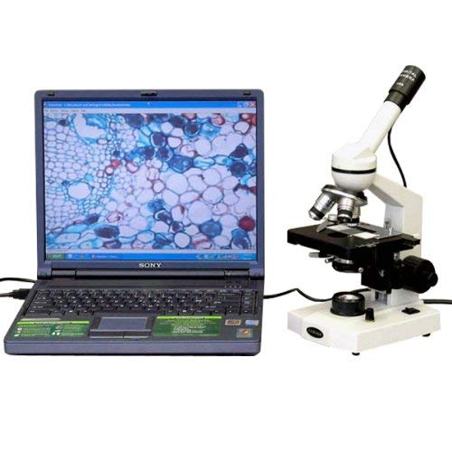
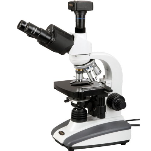
The AmScope M220C-E2 digital monocular compound microscope has interchangeable 10x and 25x widefield eyepieces, a forward-facing nosepiece with three DIN achromatic objectives, tungsten illumination, coaxial nested coarse and fine focus, a 1.25 NA Abbe condenser, and a mechanical stage. The 2MP camera has a CMOS color sensor, image capture and editing software, and USB 2.0 output to capture or display still or video images on a computer or projector. The monocular viewing head has a fixed 45-degree vertical inclination to reduce eye and neck strain, and 360-degree rotation capability to enable sharing. The forward-facing revolving nosepiece has 4x, 10x, and 40xS DIN achromatic objectives that provide color correction of magnified images. The 40xS objective is spring loaded to prevent damage to the slide or objective when focusing. A digital compound microscope is used for inspection and dissection of specimens when two-dimensional images are desired, and where image capture, detailed records, or documentation is required.
The 2MP digital camera has a CMOS color sensor for displaying still microscopy images and streaming live videos to a computer or projector, and 40x magnification. The camera can be mounted in any 23mm eye tube. The camera includes image capture and editing software that provides still image and live video capture and editing capability, including measurement functions. The software supports JPG, TIF, GIF, PSD, WMF, and BMP file formats and is compatible with Windows XP, Vista, 7, and 8; Mac OS X; and Linux. Camera drivers are compatible with Windows XP, Vista, 7, and 8; Mac OS X; and Linux. The software includes Windows APIs for native C/C++, C#, DirectShow, Twain, and LabVIEW that enable custom application development. The camera has a USB 2.0 data port (cable included).
The microscope has lower (diascopic) Brightfield illumination that transmits light up through the specimen for enhanced visibility of translucent and transparent objects. Brightfield (BF) illumination allows the specimen to absorb light, resulting in a dark image on a light background. Tungsten (incandescent) illumination provides bright light, and a rheostat controls the amount of light emanating from the lamp. The 1.25 NA Abbe condenser can be adjusted to control the distance of the light from the stage and has an iris diaphragm to optimize the amount of light illuminating the specimen. The mechanical stage, with 1mm stage divisions, locks the slide into place and provides precise slide manipulation along the X- and Y-axis to allow coordinates to be recorded, enabling the viewer to return to a specific location on the slide. The stage is 5 x 4-1/2 inches (W x D), has a focusing range of 13/32″ (10mm), and a traveling range of 2-3/4 x 3/4 inches (W x D; where W is width, the horizontal distance from left to right; D is depth, the horizontal distance from front to back). Nested coaxial coarse and fine focus eases focusing for left- and right-handed viewers, and a rack-and-pinion mechanism provides precise and secure focusing. All the mechanical parts of the microscope are constructed of metal to provide durability and resistance to wear. The cast-alloy metal frame has a stain-resistant enamel finish for durability and to ease cleaning.
| Microscope Specifications |
| Head |
Monocular |
| Eyepieces |
WF10x, WF25x |
| Lenses |
10x, 20x, 40xS DIN achromatic |
| Stage |
Double-layer mechanical, with 1mm stage divisions |
| Focus |
Coaxial coarse and fine |
| Condenser |
1.25 NA Abbe |
| Light source |
Tungsten with rheostat, 20W |
| Diaphragm |
Iris |
| Illumination type |
Brightfield (BF) |
| Power |
110V |
| Camera Specifications |
| Resolution |
2MP (1600 x 1200 effective pixels) |
| Image type |
Still image and video display and capture |
| Camera type |
Brightfield |
| Camera sensor |
CMOS (color) |
| Magnification |
40x |
| Reduction lens |
None |
| Mounting size |
23mm |
| Computer connection |
USB 2.0 |
| File formats |
JPG, TIF, GIF, PSD, WMF, BMP |
| Software package |
Image capture and editing for Windows XP, Vista, 7, and 8; Mac OS X; and Linux |
| Camera driver compatibility |
Windows XP, Vista, 7, and 8; Mac OS X; and Linux |
Microscopes are instruments used to enhance the resolution of an object or image. Types include compound, stereo, or digital. Compound microscopes use a compound optical system with an objective lens and an eyepiece. Stereo microscopes show object depth in a three-dimensional image. Digital microscopes are used to display an image on a monitor, rather than looking through a lens. Microscopes can have monocular (one), binocular (two), or trinocular (three) eyepieces, with varying magnification abilities. Magnification ability refers to the size of an image. Resolution, also known as resolvant power, refers to the clarity of the image. The interaction between field of view (FOV), numerical aperture (NA), and working distance (WD) determines resolution. Microscopes can control magnification through a fixed focus, or through a range of adjustments. They can also utilize LED, fluorescent, and mirror light sources to help control viewing capabilities. Microscopes are widely used in education, lab research, biology, metallurgy, engineering, chemistry, manufacturing, and in the medical, forensic science, and veterinary industries.
United Scope manufactures microscopy equipment and accessories under the brand name AmScope. The company, founded in 1996, is headquartered in Irvine, CA.
What’s in the Box?
- AmScope M220C-E2 microscope with mechanical stage
- WF10x eyepiece
- WF25x eyepiece
- 4x DIN achromatic objective
- 10x DIN achromatic objective
- 40xS DIN achromatic objective
- 2MP digital camera (MD200)
- USB 2.0 cable
- Software CD
- (2) Stage clips
- Spare tungsten bulb
- Spare fuse
- Dust cover
- Instructions