AmScope SW-2T24X Trinocular Stereo Microscope, WH10x Eyepieces, 10X/20X/40X Magnification, 2X/4X Objective, Upper and Lower Halogen Lighting, Pillar Stand, 110V-120V, Includes 0.5x Barlow Lens Review

AmScope SW-2T24X Trinocular Stereo Microscope, WH10x Eyepieces, 10X/20X/40X Magnification, 2X/4X Objective, Upper and Lower Halogen Lighting, Pillar Stand, 110V-120V, Includes 0.5x Barlow Lens

    This fits your .
  • Make sure this fits by entering your model number.
  • Stereo microscope for home or classroom use enables students and hobbyists to inspect biological specimens, rocks, stamps, jewelry, and large specimens that require handling or manipulation
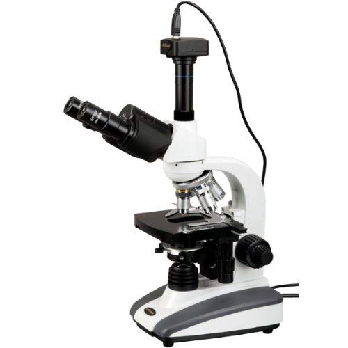
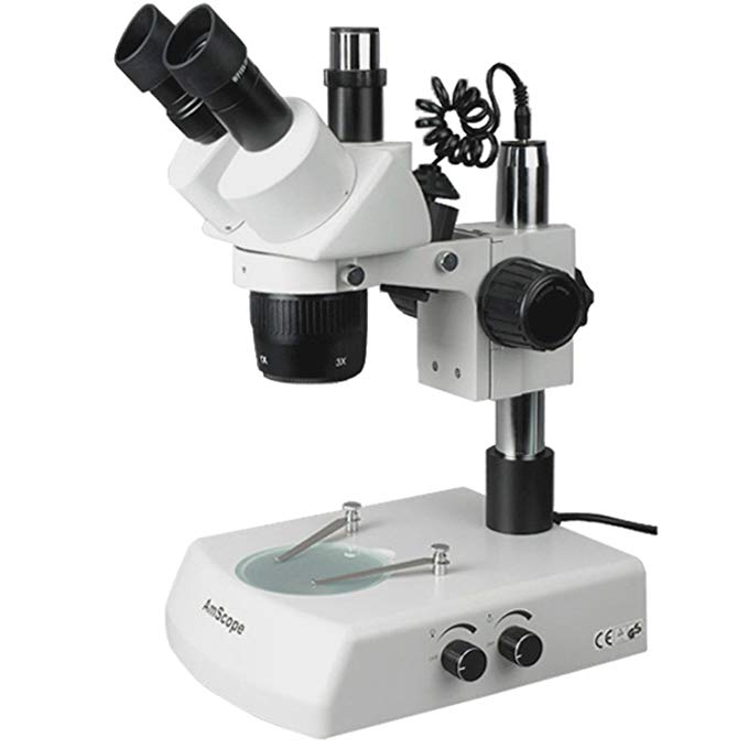
  • Trinocular viewing head with pair of 10x super-widefield high-eyepoint eyepieces, adjustable interpupillary distance, and 45-degree inclination to simplify viewing for young users
  • 2x/4x objective provides low magnification and longer focal length for inspecting large-scale specimens, and a 0.5x Barlow lens extends the working distance
  • Rheostat-controlled upper and lower illumination has 15W halogen light source to illuminate specimens, frosted stage plate allows light to pass through from below with transparent specimens, and reversible black and white stage plate provides contrast with light- and dark-colored specimens
  • Pillar-style table stand with bilateral coarse focus and stage clips to secure the specimen during viewing

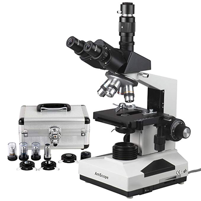
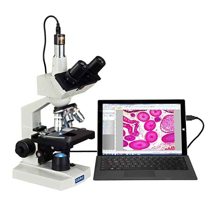
The AmScope SW2T24X trinocular stereo microscope head has a pair of 10x20mm widefield high-eyepoint eyepieces, a 2x/4x objective, and a 0.5x Barlow lens. The microscope head has a 3″ O-ring style mounting ring. The trinocular viewing head has an interpupillary range of 47 to 75mm, a 45-degree inclination to reduce eye and neck strain, and 360-degree rotation to enable sharing. The vertical trinocular port can be used as a C-Mount or 23mm photo port (camera sold separately). The WF10x20mm super-widefield high-eyepoint eyepieces combine with the 2x/4x objective to provide 10x, 20x, and 40x magnification, and a longer working distance for inspecting large-scale specimens that require handling or repair. The microscope includes a 0.5x Barlow lens that can be added to the objective to decrease the magnification range. A Barlow lens with a magnification of less than 1.0 reduces magnification and increases the working distance. High-eyepoint eyepieces ease viewing for users who wear glasses, dioptric adjustment accommodates individual eye-strength differences, and eye guards ensure comfortable viewing. The optical glass lenses provide sharp images, and are fully-coated to ensure high-resolution images. A stereo microscope, sometimes called an inspection or dissection microscope, has low magnification and a long working distance that enables users to manipulate the object being inspected.

The microscope has rheostat-controlled upper (episcopic) illumination that reflects light off the specimen for enhanced visibility of opaque specimens, and lower (diascopic) illumination that transmits light up through the specimen for enhanced visibility of translucent and transparent objects. A rheostat controls light intensity. A frosted stage plate allows light to pass through from below with transparent specimens, a reversible black and white stage plate provides contrast with light- and dark-colored specimens, and stage clips secure the specimen during viewing. The 15W halogen light source provides bright light in a concentrated path. The pillar-style stand has bilateral coarse focus to ease use for left- and right-handed users. All metal construction is durable and the enamel finish is stain-resistant.

Specifications
Head Trinocular
Magnification 10x, 20x, 40x
Objective power 2x/4x
Eyepieces (30mm) WH10x20mm
Trinocular port C-Mount or 23mm
Optical working distance Up to 4″
Field of view 0.787″ at 10x
Stand type Pillar
Illumination type Episcopic (reflected) and diascopic (transmitted)
Light source Halogen, 15W
Power 110V-120V

Microscopes are instruments used to enhance the resolution of an object or image. Types include compound, stereo, or digital. Compound microscopes use a compound optical system with an objective lens and an eyepiece. Stereo microscopes show object depth in a three-dimensional image. Digital microscopes are used to display an image on a monitor, rather than looking through a lens. Microscopes can have monocular (one), binocular (two), or trinocular (three) eyepieces, with varying magnification abilities. Magnification ability refers to the size of an image. Resolution, also known as resolvant power, refers to the clarity of the image. The interaction between field of view (FOV), numerical aperture (NA), and working distance (WD) determines resolution. Microscopes can control magnification through a fixed focus, or through a range of adjustments. They can also utilize LED, fluorescent, and mirror light sources to help control viewing capabilities. Microscopes are widely used in education, lab research, biology, metallurgy, engineering, chemistry, manufacturing, and in the medical, forensic science, and veterinary industries.

United Scope manufactures microscopy equipment and accessories under the brand name AmScope. The company, founded in 1996, is headquartered in Irvine, CA.

What’s in the Box?

  • Trinocular stereo zoom head with 2x/4x objective
  • WH10x eyepieces, one pair
  • 0.5x Barlow lens
  • Dual-illumination pillar stand with focus rack
  • Stage clips, one pair
  • Eye guards, one pair

OMAX 40X-2000X Lab Trinocular Compound LED Microscope with 1.3MP Camera Review

OMAX 40X-2000X Lab Trinocular Compound LED Microscope with 1.3MP Camera

    This fits your .
  • Make sure this fits by entering your model number.
  • Total magnification: 40X-80X-100X-200X-400X-800X-1000X-2000X; Eyepieces: wide field WF10X and WF20X; Objective: achromatic DIN 4X, 10X, 40X(S), 100X(S, Oil); Viewing head: 45 degrees inclined 360 degrees swiveling trinocular; Sliding adjustable interpupillary distance: 2-3/16inch ~ 2-15/16inch(55mm ~ 75mm)
  • Ocular diopter adjustable on both eyetubes; Nosepiece: revolving quadruple; Stage: mechanical double layer size: 4-1/2inchx 4-15/16inch (115mm x 125mm); Stage x-y stroke (travel range): 2-13/16inch x 1-3/16inch (70mm x 30mm); Condenser and diaphragm: NA1.25 Abbe condenser with iris diaphragm
  • Transmitted (lower) illuminator: LED light, intensity adjustable; Focus adjustment: Coaxial coarse and fine knobs on both sides; All metal mechanical components; Power supply: AC/DC adapter, 100V-240V (UL approved)
  • Digital camera: – True color 1280×1024 pixels (1.3MP) – Frame speed: 15fps at 1280×1024, 26fps at 640×512, 50fps at 320×256 – Software compatible with Windows and Mac OS X operating systems – Offers capturing still images, stitching, EDF, video recording and measurement functions – USB cable included
  • 5-year warranty against manufacturing defects

This is a digital biological trinocular compound microscope that includes a digital imaging system. The trinocular microscopy comes with a 45° inclined 360° swiveling trinocular sliding viewing head, two pairs of widefield eyepieces, four achromatic objectives, stain-resistant double layer mechanical stage with scales, coarse & fine focusing, NA 1.25 Abbe condenser and variable intensity LED transmitted illumination system. It offers eight levels of magnification power: 40X-80X-100X-200X-400X-800X-1000X-2000X. The 1.3MP (1280×1024 pixels) USB digital imaging system captures still microscope images and live video on computer. The included software compatible with Windows, and Mac OS X (see version information below). The user-friendly editing and processing software for Windows offers advanced features including stitching, EDF (Extended Depth of Focus), video recording and measurement functions. The software allows you to make notes, shapes, and watermarks with ease. You can analyze, present, and share images of samples and specimens using the software. This type of digital compound microscopy is perfect for teaching demonstration and clinical examination, laboratories and advanced applications.

AmScope T490A-PCS Compound Trinocular Microscope, WF10x and WF16x Eyepieces, 40X-1600X Magnification, Brightfield, Halogen Illumination, Abbe Condenser, Double-Layer Mechanical Stage, Sliding Head, High-Resolution Optics, Includes 3 Phase-Contrast Objectives and Rings Review

AmScope T490A-PCS Compound Trinocular Microscope, WF10x and WF16x Eyepieces, 40X-1600X Magnification, Brightfield, Halogen Illumination, Abbe Condenser, Double-Layer Mechanical Stage, Sliding Head, High-Resolution Optics, Includes 3 Phase-Contrast Objectives and Rings

    This fits your .
  • Make sure this fits by entering your model number.
  • Trinocular compound microscope provides high magnification for biological use and educational applications
  • Trinocular viewing head with vertical camera mount and Siedentopf binocular mount with interchangeable pairs of 10x widefield and 16x widefield eyepieces and sliding head with 55 to 75mm interpupillary adjustment, fixed 30-degree vertical inclination to reduce eye and neck strain, and 360-degree rotation capability to provide a more comprehensive view and enable sharing
  • Forward-facing nosepiece with 4x, 10x, 40xS (spring), and 100xS (spring, oil) DIN achromatic objectives that provide color correction of magnified images; PH10x, PH40x, and PH100x phase-contrast plan objectives for simple phase-contrast viewing
  • Brightfield, halogen illumination with rheostat to control light intensity, and 1.25 NA Abbe condenser with iris diaphragm and rack-and-pinion focus control for precise focus, clear examination, and light control
  • Double-layer mechanical stage with 1mm stage divisions and stage stop; graduated coarse and fine focus with tension control to prevent stage drift

The AmScope T490A-PCS compound trinocular microscope has a sliding head, interchangeable pairs of 10x widefield and 16x widefield eyepieces, a forward-facing nosepiece with four DIN achromatic objectives, three phase-contrast objectives, Brightfield and phase-contrast halogen illumination, and a double-layer mechanical stage with a stage stop to protect slides and objectives from damage. The trinocular viewing head has a vertical camera mount and a compensation-free binocular viewing head with interchangeable pairs of WF10x and WF16x eyepieces and sliding head to adjust inter-pupillary distance, a fixed 30-degree vertical inclination to reduce eye and neck strain, and a 360-degree rotation capability to provide a more comprehensive view and enable sharing. A Siedentopf binocular head enables the viewer to change the interpupillary distance without changing the tube length, eliminating the need to re-focus the image. It has an interpupillary range of 55 to 75mm to accommodate individual eye differences, and bi-lateral dioptric adjustment accommodates individual eye-strength differences. The vertical trinocular port accepts a camera with a 23mm or C-Mount adapter (camera sold separately). The forward-facing nosepiece has 4x, 10x, 40xS (spring), and 100xS (spring, oil) DIN achromatic optical-glass objectives that combine with the eyepieces to provide color correction of magnified images. The 40xS objective is spring loaded to prevent slide damage when focusing. The 100xS spring-loaded oil-immersion objective uses oil between the specimen and the objective lens to provide increased resolution over a standard objective. Three phase-contrast plan objectives (PH10x, PH40x, and PH100x) combine with the eyepieces to provide phase contrast at six magnifications. Plan objectives provide improved focus over the entire range of the viewing field. A compound microscope is used for inspection and dissection of specimens when two-dimensional images are desired.

The microscope has lower (transmitted, diascopic) Brightfield and phase-contrast illumination that transmits light up through the specimen for enhanced visibility of translucent and transparent objects. Brightfield (BF) illumination allows the specimen to absorb light, resulting in a dark image on a light background. Phase-contrast provides high contrast and visibility without the use of stains, allowing specimens to be observed in their natural state without being killed or fixed. Halogen illumination provides bright light in a concentrated path and has a rheostat to control light intensity. The 1.25 NA Abbe condenser is mounted on a rack-and-pinion control system, can be adjusted to control the distance of the light from the stage, and has an iris diaphragm to optimize the amount of light illuminating the specimen. The double-layer mechanical stage, with 1mm stage divisions, locks the slide into place and provides precise slide manipulation along the X- and Y-axis to allow coordinates to be recorded, enabling the viewer to return to a specific location on the slide. A stage stop prevents the stage from coming into contact with the slide and objectives. The stage is 5-1/2 x 5-1/2 inches (140 x 140mm) and has a traveling range of 2-15/16 x 1-3/4 inches (75 x 45mm). Graduated coaxial coarse and fine focus has a focusing range of 1-3/16″ (30mm) and fine-focus divisions of 0.002mm (0.0000787″) that enable measurements to be taken. Focus knob tension control prevents the stage from drifting out of focus. The solid-metal enamel-coated body is durable and resistant to stains and corrosion.

Specifications
Head Compound trinocular
Magnification 40x-1600x
Trinocular port 23mm or C-Mount
Eyepieces WF10x, WF16x
Objectives 4X, 10x, 40xS, 100xS (oil), PH10x, PH40x, PH100x
Stage Double-layer mechanical
Lighting configuration Transmitted (lower)
Condenser 1.25 NA Abbe
Diaphragm Iris
Light source Halogen with rheostat, 6V/20W
Illumination type Brightfield/Phase-contrast
Power 110V

Microscopes are instruments used to enhance the resolution of an object or image. Types include compound, stereo, or digital. Compound microscopes use a compound optical system with an objective lens and an eyepiece. Stereo microscopes show object depth in a three-dimensional image. Digital microscopes are used to display an image on a monitor, rather than looking through a lens. Microscopes can have monocular (one), binocular (two), or trinocular (three) eyepieces, with varying magnification abilities. Magnification ability refers to the size of an image. Resolution, also known as resolvant power, refers to the clarity of the image. The interaction between field of view (FOV), numerical aperture (NA), and working distance (WD) determines resolution. Microscopes can control magnification through a fixed focus, or through a range of adjustments. They can also utilize LED, fluorescent, and mirror light sources to help control viewing capabilities. Microscopes are widely used in education, lab research, biology, metallurgy, engineering, chemistry, manufacturing, and in the medical, forensic science, and veterinary industries.

United Scope manufactures microscopy equipment and accessories under the brand name AmScope. The company, founded in 1996, is headquartered in Irvine, CA.

What’s in the Box?

  • AmScope T490A-PCS microscope with double-layer mechanical stage
  • WF10x eyepieces, 23mm, one pair
  • WF16x eyepieces, 23mm, one pair
  • 4x DIN achromatic objective
  • 10x DIN achromatic objective, 20mm
  • 40xS DIN achromatic objective, 20mm
  • 100xS (oil) DIN achromatic objective, 20mm
  • PH10x (0.25) phase-contrast objective and condenser
  • PH40x (0.65) phase-contrast objective and condenser
  • PH100x (1.25) phase-contrast objective and condenser
  • Immersion oil, one bottle
  • (3) Color filters; blue, green, and yellow
  • Spare halogen bulb
  • Spare fuse
  • Dust cover
  • Power cord
  • Instructions

AmScope M500B-MS-M Digital Monocular Compound Microscope, WF10x and WF20x Eyepieces, 40x-2000x Magnification, Anti-Mold Optics, Tungsten Illumination, Brightfield, Abbe Condenser, Coarse and Fine Focus, Plain Stage with Mechanical Specimen Holder, 110V, Includes 1.3MP Camera with Reduction Lens and Software Review

AmScope M500B-MS-M Digital Monocular Compound Microscope, WF10x and WF20x Eyepieces, 40x-2000x Magnification, Anti-Mold Optics, Tungsten Illumination, Brightfield, Abbe Condenser, Coarse and Fine Focus, Plain Stage with Mechanical Specimen Holder, 110V, Includes 1.3MP Camera with Reduction Lens and Software

    This fits your .
  • Make sure this fits by entering your model number.
  • Digital compound microscope and a 1.3MP camera with reduction lens and USB 2.0 output for capturing or displaying images on a computer or projector
  • Monocular viewing head with interchangeable 10x and 20x widefield eyepieces, fixed 45-degree vertical inclination to reduce eye and neck strain, 360-degree rotation capability to enable sharing, and anti-mold head to preserves optics in high-humidity area
  • Forward-facing nosepiece with precision-ground 4x, 10x, 40xS, and 100xS (oil) DIN achromatic glass objectives provide color correction of magnified images
  • Brightfield imaging with rheostat-controlled tungsten (incandescent) illumination and spiral-control 1.25 NA Abbe condenser with iris diaphragm and color filter
  • Plain stage with clips to secure specimen and mechanical specimen holder to provide precise movement and record measurements; coaxial coarse and fine focus on rack-and-pinion mechanism to provide precise focus, and tension control to prevent stage drift

The AmScope M500B-MS-M digital monocular compound microscope has interchangeable 10x and 20x widefield eyepieces, a forward-facing nosepiece with four DIN achromatic objectives, tungsten illumination, coaxial separate coarse and fine focus, a 1.25 NA Abbe condenser, and a plain stage with a mechanical specimen holder. The 1.3MP camera has a CMOS color sensor, a reduction lens, image capture and editing software, and USB 2.0 output to capture or display still or video images on a computer or projector. The monocular viewing head has a fixed 45-degree vertical inclination to reduce eye and neck strain, and 360-degree rotation capability to enable sharing. An anti-mold head preserves optics in high-humidity areas. The forward-facing revolving nosepiece has 4x, 10x, 40xS, and 100xS (oil) DIN achromatic objectives that provide color correction of magnified images. The 40xS and 100xS objectives are spring loaded to prevent damage to the slide or objective when focusing. The 100xS oil-immersion objective uses oil between the specimen and the objective lens to provide increased resolution over a standard objective. The fully-coated optical system provides sharp, high-resolution images. A digital compound microscope is used for inspection and dissection of specimens when two-dimensional images are desired, and where image capture, detailed records, or documentation is required.

The 1.3MP digital camera has a CMOS color sensor for displaying still microscopy images and streaming live videos to a computer or projector. The camera has 40x magnification and a 0.37x reduction lens that ensures that the display has a similar field of view as the microscope eyepiece. The camera can be mounted in a C-Mount or any 23mm eye tube. The camera includes image capture and editing software that provides still image and live video capture and editing capability, including measurement functions. The software supports JPG, TIF, GIF, PSD, WMF, and BMP file formats and is compatible with Windows XP, Vista, 7, and 8; Mac OS X; and Linux. Camera drivers are compatible with Windows XP, Vista, 7, and 8; Mac OS X; and Linux. The software includes Windows APIs for native C/C++, C#, DirectShow, Twain, and LabVIEW that enable custom application development. The camera has a USB 2.0 data port (cable included).

The microscope has lower (diascopic) Brightfield illumination that transmits light up through the specimen for enhanced visibility of translucent and transparent objects. Brightfield (BF) illumination allows the specimen to absorb light, resulting in a dark image on a light background. Tungsten (incandescent) illumination provides bright light, and a rheostat controls the amount of light emanating from the lamp. The 1.25 NA Abbe condenser can be adjusted to control the distance of the light from the stage and has an iris diaphragm to optimize the amount of light illuminating the specimen. The condenser is controlled using a spiral mechanism. The plain stage has clips to secure the specimen in place, and is 4-3/8 x 4-3/4 inches (110 x 120mm) (W x D; where W is width, the horizontal distance from left to right; D is depth, the horizontal distance from front to back). A mechanical specimen holder can be added to the plain stage to provide precise movement, perform measurements, and record coordinates. Separate coaxial coarse and fine focus eases focusing for left- and right-handed viewers, and a rack-and-pinion mechanism provides precise and secure focusing. Focus tension control prevents stage drift. All the mechanical parts of the microscope are constructed of metal to provide durability and resistance to wear. The metal frame has a stain-resistant enamel finish for durability and to ease cleaning.

Microscope Specifications
Head Monocular
Eyepieces WF10x, WF20x (23mm)
Lenses 10x, 20x, 40xS, 100xS (oil) DIN achromatic (20mm)
Stage Plain stage with stage clips and mechanical specimen holder
Focus Coaxial coarse and fine
Condenser 1.25 NA Abbe
Light source Tungsten with rheostat, 20W
Diaphragm Iris
Illumination type Brightfield (BF)
Power 110V

Camera Specifications
Resolution 1.3MP (1280 x 1024 effective pixels)
Image type Still image and video display and capture
Camera type Brightfield
Camera sensor 1/2″ Aptina MT9M111 CMOS (color)
Magnification 40x
Reduction lens 0.37x
Mounting size 23mm or C-Mount
Frame rate 50fps at 320×256; 26fps at 640×512; 15fps at 1280×1024
Computer connection USB 2.0 (backward compatible on PCs only)
File formats JPG, TIF, GIF, PSD, WMF, BMP
Software package Image capture and editing for Windows XP, Vista, 7, and 8; Mac OS X; and Linux
Camera driver compatibility Windows XP, Vista, 7, and 8; Mac OS X; and Linux

Microscopes are instruments used to enhance the resolution of an object or image. Types include compound, stereo, or digital. Compound microscopes use a compound optical system with an objective lens and an eyepiece. Stereo microscopes show object depth in a three-dimensional image. Digital microscopes are used to display an image on a monitor, rather than looking through a lens. Microscopes can have monocular (one), binocular (two), or trinocular (three) eyepieces, with varying magnification abilities. Magnification ability refers to the size of an image. Resolution, also known as resolvant power, refers to the clarity of the image. The interaction between field of view (FOV), numerical aperture (NA), and working distance (WD) determines resolution. Microscopes can control magnification through a fixed focus, or through a range of adjustments. They can also utilize LED, fluorescent, and mirror light sources to help control viewing capabilities. Microscopes are widely used in education, lab research, biology, metallurgy, engineering, chemistry, manufacturing, and in the medical, forensic science, and veterinary industries.

United Scope manufactures microscopy equipment and accessories under the brand name AmScope. The company, founded in 1996, is headquartered in Irvine, CA.

What’s in the Box?

  • AmScope M500B-MS-M microscope with plain stage and mechanical specimen holder
  • WF10x eyepiece, 23mm
  • WF20x eyepiece, 23mm
  • 4x DIN achromatic objective, 20mm
  • 10x DIN achromatic objective, 20mm
  • 40xS DIN achromatic objective, 20mm
  • 100xS (oil) DIN achromatic objective, 20mm
  • Mechanical specimen holder
  • 1.3MP digital camera (MU130)
  • 0.37x reduction lens
  • Software CD
  • USB 2.0 cable
  • (2) Stage clips
  • Wrench
  • Color filter
  • Immersion oil
  • Spare bulb
  • Dust cover
  • Instructions

OMAX 40X-2500X USB3 10MP Digital Darkfield Lab Siedentopf Trinocular LED Microscope for Live Blood Review

OMAX 40X-2500X USB3 10MP Digital Darkfield Lab Siedentopf Trinocular LED Microscope for Live Blood

    This fits your .
  • Make sure this fits by entering your model number.
  • Total magnification: 40X-100X-250X-400X-1000X-2500X; Eyepieces: wide field WF10X/18 and WF25X; Objective: achromatic DIN 4X, 10X, 40X (spring),100X (spring, oil); Head: 30 degrees inclined 360 degrees rotatable trinocular siedentopf; Interpupillary: hinge adjustment 1-7/8inch ~ 2-15/16inch (48mm ~ 75mm); Diopter: adjustable on left eyepiece tube; Nosepiece: revolving, quadruple, reversed; Stage: X-Y mechanical stage with scales, range 78mm x 40mm, dimension 110mm x 140mm
  • Focus: coaxial coarse and fine focus knobs on both sides, rack and pinion adjustment, with tension control and stop; Illumination: transmitted replaceable 3W LED light, intensity adjustable; Brightfield condenser: NA1.25 Abbe condenser with iris diaphragm and filter holder, rack and pinion adjustment; Oil darkfield condenser: NA1.36-1.25 condenser plus plan 100X/1.25-0.5 160/0.17 objective (oil, spring) with built-in iris diaphragm
  • Solid metal base and stand; Power supply: AC 100V ~240V 50/60Hz (US and Canada plug)
  • USB3 Digital camera – true color 3584×2748 pixels (10M pixels) – 0.5X reduction lens to get larger field of view – 0.01 mm calibration slide: 1mm/100 division – Frame speed: 7.2fps at 3584×2746, 25fps at 1792×1372 – Software compatible with Windows XP/Vista/7/8/10, Mac OS X and Linux (2.6 and later) operating systems – Capturing microscope images, recording live video, measuring lengths, angles, areas, editing images – USB3 cable included
  • 5-year warranty against manufacturing defects

This trinocular compound LED siedentopf microscope 40X-2500X provides brightfield & darkfield microscope system and utilizes a super speed 10MP USB 3.0 digital imaging system. The darkfield microscope features an optical system for simultaneously observing images through eyepieces and an adjustable trinocular port. The 3584×2748 pixels USB 3.0 digital imaging system that captures still microscope images and live video on computer. The included software compatible with windows, Mac OS and Linux operating system can also be used to make measurements. All lenses are made of high quality optical glass. The high resolution image in the field of view is crystal clear and will satisfy the demands of professionals. This darkfield compound microscopy is ideal for professionals and also affordable for students.

AmScope M60A-BTK Beginner Microscope Kit, Mirror Illumination, WF10x and WF16x Eyepieces, 40x-640x Magnification, Includes Case, 5 Blank Slides, 5 Prepared Slides, and Book Review

AmScope M60A-BTK Beginner Microscope Kit, Mirror Illumination, WF10x and WF16x Eyepieces, 40x-640x Magnification, Includes Case, 5 Blank Slides, 5 Prepared Slides, and Book

    This fits your .
  • Make sure this fits by entering your model number.
  • Beginner compound microscope provides high magnification for biological use and educational applications
  • Monocular viewing head with 10x widefield and 16x widefield eyepieces and a fixed upright inclination
  • Forward-facing nosepiece with 4x, 10x, and 40x DIN achromatic objectives that provide color correction of magnified images
  • Brightfield, mirror illumination, and 0.65 NA single-lens condenser with disc diaphragm
  • Plain stage with stage clips to secure slide and a stage stop to prevent damage to slides or objectives

The AmScope M60A-BTK monocular compound microscope has 10x widefield and 16x widefield eyepieces, a forward-facing nosepiece with three DIN achromatic objectives, Brightfield mirror illumination, a 0.65 NA single-lens condenser, and a plain stage. The forward-facing revolving nosepiece has 4x, 10x, and 40x DIN achromatic objectives that provide color correction of magnified images. The monocular viewing head holds the WF10x or WF16x eyepiece on a fixed upright inclination. A compound microscope is used for inspection and dissection of specimens when two-dimensional images are desired.

The microscope has lower (diascopic) Brightfield illumination that transmits light up through the specimen for enhanced visibility of translucent and transparent objects. Brightfield (BF) illumination allows the specimen to absorb light, resulting in a dark image on a light background. A mirror directs light onto the specimen without the need for power or batteries. The 0.65 NA single-lens condenser and disc diaphragm control the amount of light illuminating the specimen. The plain stage has an opening for light transmission and stage clips to secure the slide or specimen in place while viewing. A stage stop prevents the stage or specimen from coming into contact with the objectives. The solid-metal frame has a stain-resistant enamel finish for durability, and a hard-sided plastic case protects the microscope during storage or transport. The microscope comes with five blank slides, five prepared slides, and the World of the Microscope book.

Specifications
Head Compound monocular
Eyepieces WF10x, WF16x
Objectives 4X, 10x, 40x DIN achromatic
Stage Plain
Lighting configuration Transmitted (lower)
Condenser Single lens
Diaphragm Disc diaphragm
Light source Mirror
Illumination type Brightfield
Power N/A

Microscopes are instruments used to enhance the resolution of an object or image. Types include compound, stereo, or digital. Compound microscopes use a compound optical system with an objective lens and an eyepiece. Stereo microscopes show object depth in a three-dimensional image. Digital microscopes are used to display an image on a monitor, rather than looking through a lens. Microscopes can have monocular (one), binocular (two), or trinocular (three) eyepieces, with varying magnification abilities. Magnification ability refers to the size of an image. Resolution, also known as resolvant power, refers to the clarity of the image. The interaction between field of view (FOV), numerical aperture (NA), and working distance (WD) determines resolution. Microscopes can control magnification through a fixed focus, or through a range of adjustments. They can also utilize LED, fluorescent, and mirror light sources to help control viewing capabilities. Microscopes are widely used in education, lab research, biology, metallurgy, engineering, chemistry, manufacturing, and in the medical, forensic science, and veterinary industries.

United Scope manufactures microscopy equipment and accessories under the brand name AmScope. The company, founded in 1996, is headquartered in Irvine, CA.

What’s in the Box?

  • AmScope M60A microscope with plain stage
  • WF10x eyepiece
  • WF16x eyepiece
  • 4x DIN achromatic objective
  • 10x DIN achromatic objective
  • 40x DIN achromatic objective
  • (5) Blank slides
  • (5) Prepared slides
  • (2) Stage clips
  • World of the Microscope book
  • Carrying case
  • Instructions

AmScope T360B-8M Digital Trinocular Compound Microscope, 40X-2000X Magnification, WF10x and WF20x Eyepieces, Brightfield, LED Illumination, Abbe Condenser, Double-Layer Mechanical Stage, Includes 8MP Camera with Reduction Lens and Software Review

AmScope T360B-8M Digital Trinocular Compound Microscope, 40X-2000X Magnification, WF10x and WF20x Eyepieces, Brightfield, LED Illumination, Abbe Condenser, Double-Layer Mechanical Stage, Includes 8MP Camera with Reduction Lens and Software

    This fits your .
  • Make sure this fits by entering your model number.
  • Digital compound microscope and an 8MP camera with reduction lens and USB 2.0 output for capturing or displaying images on a computer or projector
  • Siedentopf trinocular viewing head with pairs of 10x and 20x widefield eyepieces and 48 to 75mm interpupillary adjustment, fixed 30-degree vertical inclination to reduce eye and neck strain, and 360-degree rotation capability to provide a more comprehensive view and enable sharing
  • Reverse-mounted nosepiece with 4x, 10x, 40xS (spring), and 100xS (spring, oil) DIN achromatic objectives that provide color correction of magnified images
  • Brightfield, LED illumination, and 1.25 NA Abbe condenser with iris diaphragm for clear examination and light control
  • Double-layer mechanical stage, with 1.0mm stage divisions, locks slide into place and provides precise slide manipulation along the X- and Y-axis to allow coordinates to be recorded, enabling the viewer to return to a specific location on the slide

The AmScope T360B-8M digital compound trinocular microscope has interchangeable pairs of 10x and 20x widefield eyepieces, a reverse-mounted nosepiece with four DIN achromatic objectives, Brightfield LED illumination, and a double-layer mechanical stage. The 8MP camera has a CMOS color sensor, a reduction lens, image capture and editing software, and USB 2.0 output to capture or display still or video images on a computer or projector. The vertical trinocular port can be used to mount the camera. The Siedentopf binocular viewing head has an adjustable 55 to 75mm interpupillary distance, a fixed 30-degree vertical inclination to reduce eye and neck strain, and a 360-degree rotation capability to provide a more comprehensive view and enable sharing. The sliding head has an adjustable interpupillary distance to accommodate individual eye differences. The reverse-mounted nosepiece has 4x, 10x, 40xS (spring), and 100xS (spring, oil) DIN achromatic objectives that combine with the eyepieces to provide color correction of magnified images at eight magnifications. The 40xS objective is spring loaded to prevent slide damage when focusing. The 100xS spring-loaded oil objective provides increased resolution over a standard objective. A reverse-mounted nosepiece keeps objectives out of the way to prevent damage and contamination, and to enable more secure slide handling. A digital compound microscope is used for inspection and dissection of specimens when two-dimensional images are desired, and where image capture, detailed records, or documentation is required.

The 8MP digital camera has a CMOS color sensor for displaying still microscopy images and streaming live videos to a computer or projector. The camera has 40x magnification and a 0.5x reduction lens that ensures that the display has a similar field of view as the microscope eyepiece. The camera can be mounted in a C-Mount or any 23mm eye tube. The camera includes image capture and editing software that provides still image and live video capture and editing capability, including measurement functions. The software supports JPG, TIF, GIF, PSD, WMF, and BMP file formats and is compatible with Windows XP, Vista, 7, and 8; Mac OS X; and Linux. Camera drivers are compatible with Windows XP, Vista, 7, and 8; Mac OS X; and Linux. The software includes Windows APIs for native C/C++, C#, DirectShow, Twain, and LabVIEW that enable custom application development. The camera has a USB 2.0 data port (cable included).

The microscope has lower (transmitted, diascopic) Brightfield illumination that transmits light up through the specimen for enhanced visibility of translucent and transparent objects. Brightfield (BF) illumination allows the specimen to absorb light, resulting in a dark image on a light background. LED illumination provides bright, cool light for working with temperature-sensitive or live specimens. The 1.25 NA Abbe condenser can be adjusted to control the distance of the light from the stage and has an iris diaphragm to optimize the amount of light illuminating the specimen. The double-layer mechanical stage, with 1mm stage divisions, locks the slide into place and provides precise slide manipulation along the X- and Y-axis to allow coordinates to be recorded, enabling the viewer to return to a specific location on the slide. Graduated coaxial coarse and fine focus enables measurements to be taken. The enamel-coated cast-steel body is durable and resistant to stains and corrosion.

Microscope Specifications
Head Compound binocular
Trinocular port 23mm or C-Mount
Eyepieces WF10x, WF20x
Objectives 4X, 10x, 40xS, 100xS (oil)
Stage Double-layer Mechanical
Lighting configuration Transmitted (lower)
Condenser 1.25 NA Abbe
Diaphragm Iris
Light source LED, 3W
Illumination type Brightfield
Power 110V

Camera Specifications
Resolution 8MP (3264 x 2448 effective pixels)
Image type Still image and video display and capture
Camera type Brightfield
Camera sensor 1/2.5″ Aptina Special CMOS (color)
Magnification 40x
Reduction lens 0.5x
Mounting size 23mm or C-Mount
Frame rate 27fps at 800×600; 8fps at 1600×1200; 1.9fps at 3264×2448
Computer connection USB 2.0 (backward compatible on PCs only)
File formats JPG, TIF, GIF, PSD, WMF, BMP
Software package Image capture and editing for Windows XP, Vista, 7, and 8; Mac OS X; and Linux
Camera driver compatibility Windows XP, Vista, 7, and 8; Mac OS X; and Linux

Microscopes are instruments used to enhance the resolution of an object or image. Types include compound, stereo, or digital. Compound microscopes use a compound optical system with an objective lens and an eyepiece. Stereo microscopes show object depth in a three-dimensional image. Digital microscopes are used to display an image on a monitor, rather than looking through a lens. Microscopes can have monocular (one), binocular (two), or trinocular (three) eyepieces, with varying magnification abilities. Magnification ability refers to the size of an image. Resolution, also known as resolvant power, refers to the clarity of the image. The interaction between field of view (FOV), numerical aperture (NA), and working distance (WD) determines resolution. Microscopes can control magnification through a fixed focus, or through a range of adjustments. They can also utilize LED, fluorescent, and mirror light sources to help control viewing capabilities. Microscopes are widely used in education, lab research, biology, metallurgy, engineering, chemistry, manufacturing, and in the medical, forensic science, and veterinary industries.

United Scope manufactures microscopy equipment and accessories under the brand name AmScope. The company, founded in 1996, is headquartered in Irvine, CA.

What’s in the Box?

  • AmScope T360B-8M microscope with double-layer mechanical stage
  • WF10x eyepiece, one pair
  • WF20x objectives, one pair
  • 4x DIN achromatic objective
  • 10x DIN achromatic objective
  • 40xS DIN achromatic objective
  • 100xS (oil) DIN achromatic objective
  • Immersion oil, one bottle
  • (2) Color filters, blue and green
  • 8MP digital camera (MU800)
  • 0.5x reduction lens
  • USB 2.0 cable
  • Software CD
  • Mirror
  • Dust cover
  • Power cord (US and Canada standard)
  • Instructions

OMAX 40X-2500X Digital Lab Trinocular Compound LED Microscope with USB Digital Camera and Double Layer Mechanical Stage Review

OMAX 40X-2500X Digital Lab Trinocular Compound LED Microscope with USB Digital Camera and Double Layer Mechanical Stage

    This fits your .
  • Make sure this fits by entering your model number.
  • Total magnification: 40X-100X-250X-400X-1000X-2500X; Eyepieces: wide field WF10X and WF25X; Objectives: achromatic DIN 4X, 10X, 40X(S), 100X(S, Oil); Viewing head: 45 degrees inclined 360 degrees swiveling trinocular
  • Sliding adjustable interpupillary distance: 2-3/16inch ~ 2-15/16inch(55~75mm); Ocular diopter adjustable on both eyetubes; Nosepiece: revolving quadruple; Stage: mechanical double layer size: 4-1/2inchx 4-15/16inch (115mm x 125mm); Stage x-y stroke (travel range): 2-13/16inch x 1-3/16inch (70mm x 30mm)
  • Condenser and diaphragm: NA1.25 Abbe condenser with iris diaphragm; Transmitted (lower) illuminator: LED light, intensity adjustable; Focus adjustment: Coaxial coarse and fine knobs on both sides; All metal mechanical components; Power supply: AC/DC adapter, 7.5V/7.5W (UL approved); – Input: 100-240V
  • Digital camera: – true color 640 x 480 pixels – 0.45X reduction lens to get larger field of view – Software compatible with Windows 2000/XP/Vista/7/8/10 and Mac OS – Capturing microscope images, recording live video, measuring lengths, angles, areas, editing images
  • 5-year warranty against manufacturing defects

This OMAX digital LED trinocular compound microscope includes a digital imaging system. The digital microscopy comes with a 45° inclined 360° swiveling trinocular sliding viewing head, two pairs of widefield eyepieces, four achromatic objectives, stain-resistant double layer mechanical stage with scales, coarse & fine focusing, NA 1.25 Abbe condenser and variable intensity LED transmitted illumination system. It offers six levels of magnification power: 40X-100X-250X-400X-1000X-2500X. The USB digital imaging system that captures microscope images and live video on computer. The included software compatible with Windows XP/Vista/7/8/10 can be used to make measurements, You can view and capture the images on iMac or Macbook with Mac OS too. It is easy to mount a darkfield condenser to switch a brightfield microscope to a darkfield microscope. This type of digital compound microscopy is perfect for teaching demonstration and clinical examination, laboratories and advanced applications.

Walter Products 3000F-100-LED Basic Monocular Compound Microscope, WF10x Eyepiece, 40x-1000x Magnification, Brightfield, LED Illumination, Abbe Condenser, Iris Diaphragm, Mechanical Stage, 110V Review

Walter Products 3000F-100-LED Basic Monocular Compound Microscope, WF10x Eyepiece, 40x-1000x Magnification, Brightfield, LED Illumination, Abbe Condenser, Iris Diaphragm, Mechanical Stage, 110V

The Walter Products 3000F-100-LED Basic Monocular Compound Microscope has a 10x widefield eyepiece with pointer, a forward-facing nosepiece with four DIN achromatic objectives that are parcentered and parfocal, LED illumination, nested coarse and fine focus, 1.25 NA Abbe condenser with iris diaphragm, and a mechanical stage. The monocular viewing head has a fixed inclination to reduce eye and neck strain, and rotation capability to enable sharing. An eyepiece pointer is used to identify features for students. A forward-facing nosepiece eases changing objectives. Achromatic objectives provide color correction of magnified images. Parcentered and parfocal objectives ensure that the image stays centered and focused when the magnification is changed. The 40xR objective is retractable and spring-loaded to prevent damage to the slide or objective when focusing. The 100xR objective is also an oil objective that provides increased resolution over a standard objective. A basic compound microscope is used in classroom, home school, and hobbyist settings for inspection and dissection of specimens when two-dimensional images are desired.

The microscope has lower (diascopic) brightfield illumination that transmits light up through the specimen for enhanced visibility of translucent and transparent objects. Brightfield (BF) illumination allows the specimen to absorb light, resulting in a dark image on a light background. The LED light source provides bright, cool light for working with temperature-sensitive or live specimens. The Abbe condenser can be adjusted to control the distance of the light from the stage and has an iris diaphragm to optimize the amount of light illuminating the specimen. The mechanical stage locks the slide into place and provides precise slide manipulation along the X- and Y-axes. Coaxial coarse and fine focus knobs are nested to speed focusing for left-and right-handed viewers. A stage stop prevents the stage or specimen from coming into contact with the objectives.

Microscope Head and Optics Specifications
Head Monocular
Magnification range 40x-1000x
Head inclination 45 degrees
Eyepiece WF10x with pointer
Nosepiece Forward-facing
Objectives, DIN achromatic, parcentered, parfocal 4x, 10x, 40xR (retractable), 100xR (oil, retractable)

Microscope Illumination and Stage Specifications
Focus type Coaxial nested coarse and fine
Condenser 1.25 NA Abbe
Light source LED
Diaphragm Iris
Illumination type Brightfield
Stage type Mechanical
Power 110V

Microscopes are instruments used to enhance the resolution of an object or image. Types include compound, stereo, or digital. Compound microscopes use a compound optical system with an objective lens and an eyepiece. Stereo microscopes show object depth in a three-dimensional image. Digital microscopes are used to display an image on a monitor, rather than looking through a lens. Microscopes can have monocular (one), binocular (two), or trinocular (three) eyepieces, with varying magnification abilities. Magnification ability refers to the size of an image. Resolution, also known as resolvant power, refers to the clarity of the image. The interaction between field of view (FOV), numerical aperture (NA), and working distance (WD) determines resolution. Microscopes can control magnification through a fixed focus, or through a range of adjustments. They can also utilize LED, fluorescent, and mirror light sources to help control viewing capabilities. Microscopes are widely used in education, lab research, biology, metallurgy, engineering, chemistry, manufacturing, and in the medical, forensic science, and veterinary industries.

Walter Products distributes microscopy and laboratory equipment and supplies. The company, founded in 1987, is headquartered in Windsor, ON, Canada.

What’s in the Box?

  • Walter Products 3000F-100-LED Basic Monocular Compound Microscope
  • WF10x eyepiece
  • 4x DIN achromatic objective
  • 10x DIN achromatic objective
  • 40xR (retractable) DIN achromatic objective
  • 100xR (oil, retractable) DIN achromatic objective
  • Dust cover
  • Instructions

National Optical 131-CLED Basic Monocular Compound Microscope, WF10x Eyepiece, 40x-400x Magnification, Brightfield, LED Illumination, Disc Diaphragm, Plain Stage, 110V Review

National Optical 131-CLED Basic Monocular Compound Microscope, WF10x Eyepiece, 40x-400x Magnification, Brightfield, LED Illumination, Disc Diaphragm, Plain Stage, 110V

The National Optical 131-CLED Basic Monocular Compound Microscope has a 10x widefield eyepiece with pointer, a forward-facing nosepiece with three DIN achromatic objectives that are parcentered and parfocal, LED illumination, separate coarse and fine focus, a disc diaphragm, and a plain stage with stage clips. The monocular viewing head has a fixed inclination to reduce eye and neck strain, and 360-degree rotation capability to enable sharing. An eyepiece pointer is used to identify features for students. A forward-facing nosepiece eases changing objectives. Rotation positions with secure, positive stops ensure accurate positioning and support a range of magnification options. Achromatic objectives provide color correction of magnified images. Parcentered and parfocal objectives ensure that the image stays centered and focused when the magnification is changed. The 40xR objective is retractable and spring-loaded to prevent damage to the slide or objective when focusing. A basic compound microscope is used in classroom, home school, and hobbyist settings for inspection and dissection of specimens when two-dimensional images are desired.

The microscope has lower (diascopic) Brightfield illumination that transmits light up through the specimen for enhanced visibility of translucent and transparent objects. Brightfield (BF) illumination allows the specimen to absorb light, resulting in a dark image on a light background. The LED light source provides bright, cool light for working with temperature-sensitive or live specimens. A disc diaphragm controls the amount of light illuminating the specimen. The plain stage has an opening for light transmission, stage clips to secure the slide or specimen in place while viewing, and pre-drilled holes that accept a mechanical stage (sold separately). Separate coarse and fine focus knobs speed focusing. A slip clutch prevents excess pressure from damaging the slides or objectives, a stage stop prevents the stage or specimen from coming into contact with the objectives, and a focus tension control prevents stage drift. The cast metal frame provides durability and has a gray enamel finish.

Microscope Head and Optics Specifications
Head Monocular
Magnification range 40x-400x
Head inclination 45 degrees
Eyepiece (18mm) WF10x
Nosepiece Forward-facing
Objectives, DIN achromatic, parcentered, parfocal 4x, 10x, 40xR (retractable)

Microscope Illumination and Stage Specifications
Focus type Separate coarse and fine
Condenser 0.65 NA
Light source LED
Diaphragm Disc
Illumination type Brightfield
Stage type Plain
Stage dimensions 110 x 120mm (W x D)
Power 110V

W is width, the horizontal distance from left to right; D is depth, the horizontal distance from front to back.

Microscopes are instruments used to enhance the resolution of an object or image. Types include compound, stereo, or digital. Compound microscopes use a compound optical system with an objective lens and an eyepiece. Stereo microscopes show object depth in a three-dimensional image. Digital microscopes are used to display an image on a monitor, rather than looking through a lens. Microscopes can have monocular (one), binocular (two), or trinocular (three) eyepieces, with varying magnification abilities. Magnification ability refers to the size of an image. Resolution, also known as resolvant power, refers to the clarity of the image. The interaction between field of view (FOV), numerical aperture (NA), and working distance (WD) determines resolution. Microscopes can control magnification through a fixed focus, or through a range of adjustments. They can also utilize LED, fluorescent, and mirror light sources to help control viewing capabilities. Microscopes are widely used in education, lab research, biology, metallurgy, engineering, chemistry, manufacturing, and in the medical, forensic science, and veterinary industries.

National Optical and Scientific Instruments manufactures microscopes and microscope cameras. The company, founded in 1991, is headquartered in Schertz, TX.

What’s in the Box?

  • National Optical 131-CLED Basic Monocular Compound Microscope
  • WF10x eyepiece, 18mm
  • 4x DIN achromatic objective
  • 10x DIN achromatic objective
  • 40xR (retractable) DIN achromatic objective
  • Hex key wrench
  • Dust cover
  • Instructions

Scroll To Top