
- Make sure this fits by entering your model number.
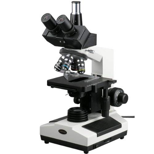
- Trinocular compound microscope provides high magnification for biological use and educational applications
- Sliding trinocular viewing head with pairs of 10x widefield and 25x widefield achromatic eyepieces and sliding head to adjust interpupillary distance, fixed 45-degree vertical inclination to reduce eye and neck strain, and 360-degree rotation capability to provide a more comprehensive view and enable sharing
- Forward-facing nosepiece with 4x, 10x, 40xS (spring), and 100xS (spring, oil) DIN achromatic objectives that provide color correction of magnified images
- Brightfield halogen illumination and 1.25 NA Abbe condenser with iris diaphragm and rack-and-pinion focus control for precise focus, clear examination, and light control
- Double-layer mechanical stage, with 1.0mm stage divisions, locks slide into place and provides precise slide manipulation along the X- and Y-axis to allow coordinates to be recorded, enabling the viewer to return to a specific location on the slide
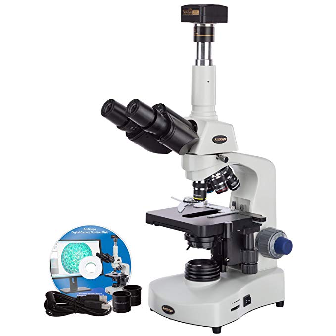
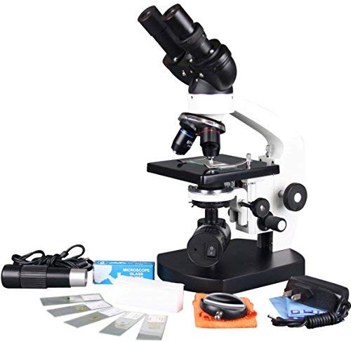
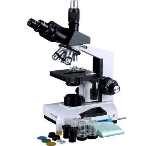
The AmScope T390C professional compound trinocular microscope interchangeable pairs of 10x widefield and 25x widefield eyepieces, a forward-facing nosepiece with four DIN achromatic objectives, Brightfield halogen illumination, and a double-layer mechanical stage. The trinocular head has a sliding binocular mount with an adjustable 55 to 75mm interpupillary distance, a fixed 30-degree vertical inclination to reduce eye and neck strain, and a 360-degree rotation capability to provide a more comprehensive view and enable sharing. Dioptric adjustment accommodates individual eye-strength differences. The vertical trinocular port accepts a camera with a 23mm or C-Mount adapter (camera sold separately). The forward-facing nosepiece has 4x, 10x, 40xS (spring), and 100xS (spring, oil) DIN achromatic objectives that combine with the eyepieces to provide color correction of magnified images at eight magnifications. The 40xS objective is spring loaded to prevent slide damage when focusing. The 100xS spring-loaded oil-immersion objective uses oil between the specimen and the objective lens to provide increased resolution over a standard objective. A compound microscope is used for inspection and dissection of specimens when two-dimensional images are desired.
The microscope has lower (transmitted, diascopic) Brightfield illumination that transmits light up through the specimen for enhanced visibility of translucent and transparent objects. Brightfield (BF) illumination allows the specimen to absorb light, resulting in a dark image on a light background. Halogen illumination provides bright light in a concentrated path. The 1.25 NA Abbe condenser is mounted on a rack-and-pinion control system, can be adjusted to control the distance of the light from the stage, and has an iris diaphragm to optimize the amount of light illuminating the specimen. The double-layer mechanical stage, with 1mm stage divisions, locks the slide into place and provides precise slide manipulation along the X- and Y-axis to allow coordinates to be recorded, enabling the viewer to return to a specific location on the slide. The stage is 5-1/2 x 5-1/2 inches (140 x 140mm) and has a traveling range of 3 x 2 inches (75 x 50mm). Graduated coaxial coarse and fine focus enables measurements to be taken. The enamel-coated cast-steel body is durable and resistant to stains and corrosion.
| Specifications | |
|---|---|
| Head | Compound trinocular |
| Trinocular port | 23mm or C-Mount |
| Eyepieces | WF10x, WF25x |
| Objectives | 4X, 10x, 40xS, 100xS (oil) |
| Stage | Double-layer mechanical |
| Lighting configuration | Transmitted (lower) |
| Condenser | 1.25 NA Abbe |
| Diaphragm | Iris |
| Light source | Halogen, 6V/20W |
| Illumination type | Brightfield |
| Power | 110V/220V, auto-switching |
| Weight | 10lb./4.5kg |
Microscopes are instruments used to enhance the resolution of an object or image. Types include compound, stereo, or digital. Compound microscopes use a compound optical system with an objective lens and an eyepiece. Stereo microscopes show object depth in a three-dimensional image. Digital microscopes are used to display an image on a monitor, rather than looking through a lens. Microscopes can have monocular (one), binocular (two), or trinocular (three) eyepieces, with varying magnification abilities. Magnification ability refers to the size of an image. Resolution, also known as resolvant power, refers to the clarity of the image. The interaction between field of view (FOV), numerical aperture (NA), and working distance (WD) determines resolution. Microscopes can control magnification through a fixed focus, or through a range of adjustments. They can also utilize LED, fluorescent, and mirror light sources to help control viewing capabilities. Microscopes are widely used in education, lab research, biology, metallurgy, engineering, chemistry, manufacturing, and in the medical, forensic science, and veterinary industries.
United Scope manufactures microscopy equipment and accessories under the brand name AmScope. The company, founded in 1996, is headquartered in Irvine, CA.
What’s in the Box?
- AmScope T390C microscope with double-layer mechanical stage
- WF10x eyepiece, one pair
- WF25x eyepiece, one pair
- 4x DIN achromatic objective
- 10x DIN achromatic objective
- 40xS DIN achromatic objective
- 100xS (oil) DIN achromatic objective
- Immersion oil, one bottle
- (3) Color filters, blue, green, and yellow
- Spare halogen bulb
- Dust cover
- Power cord (US and Canada standard)
- Instructions