AmScope T380C-M Digital Professional Compound Trinocular Microscope, 40X-1000X Magnification, WF10x and WF25x Eyepieces, Brightfield, LED Illumination, Abbe Condenser, Double-Layer Mechanical Stage, 85-265V Wide-Voltage Power Supply, Includes 1.3MP Camera with Reduction Lens and Software Review

AmScope T380C-M Digital Professional Compound Trinocular Microscope, 40X-1000X Magnification, WF10x and WF25x Eyepieces, Brightfield, LED Illumination, Abbe Condenser, Double-Layer Mechanical Stage, 85-265V Wide-Voltage Power Supply, Includes 1.3MP Camera with Reduction Lens and Software

    This fits your .
  • Make sure this fits by entering your model number.
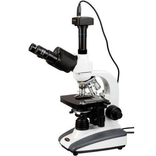
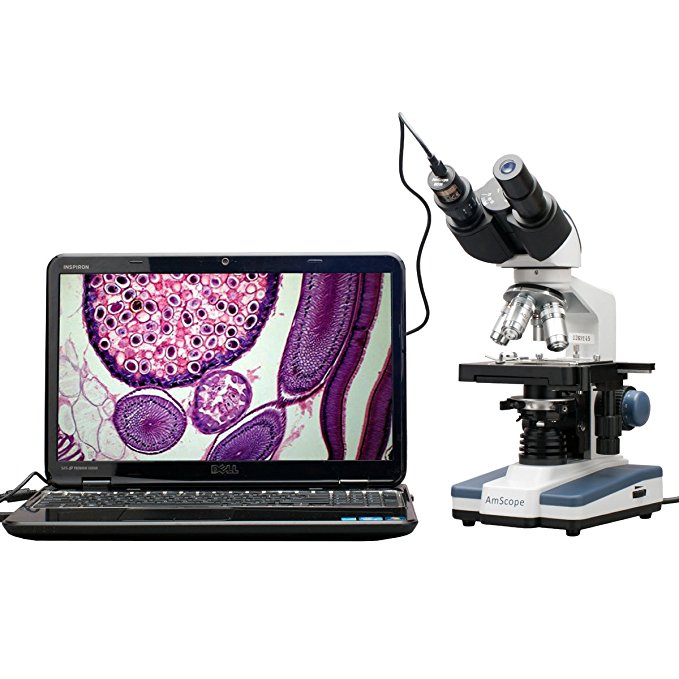
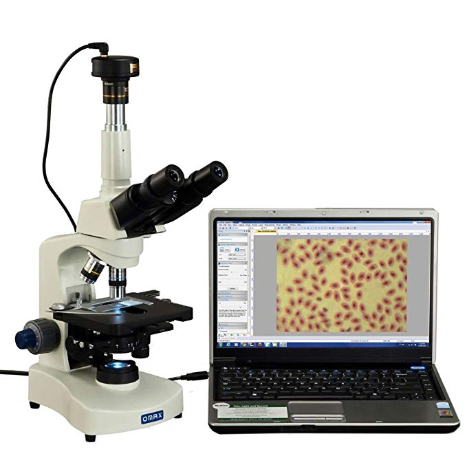
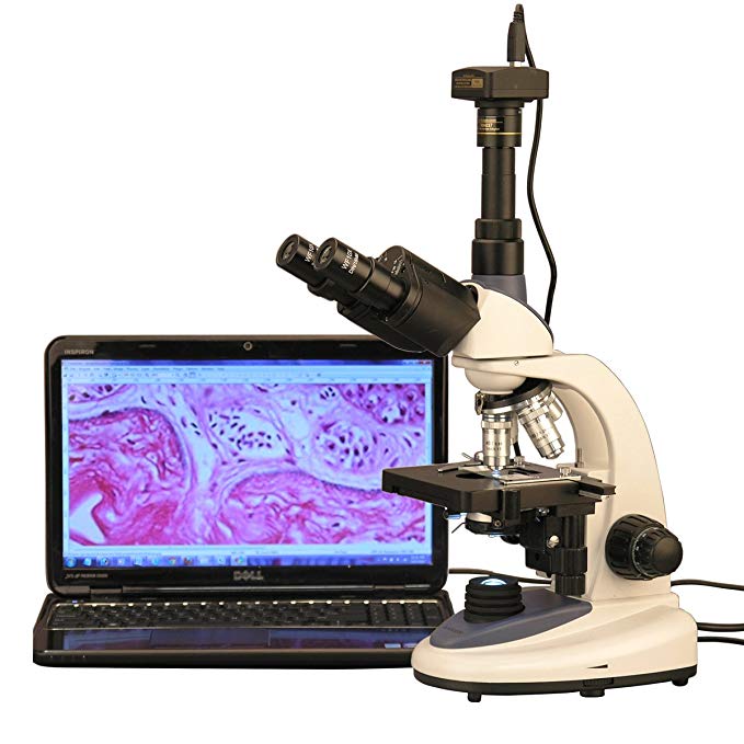
  • Digital trinocular compound microscope provides high magnification for biological use and educational applications, and has a 1.3MP camera with reduction lens and USB 2.0 output for capturing or displaying images on a computer or projector
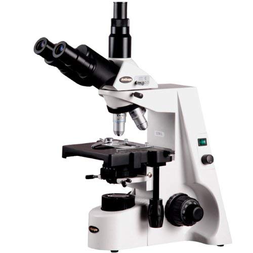
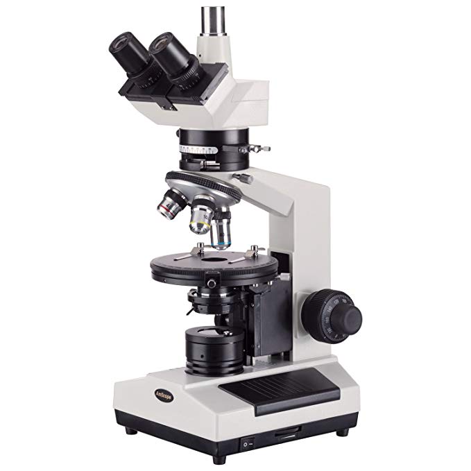
  • Siedentopf trinocular viewing head with interchangeable pairs of 10x and 25x widefield eyepieces and adjustable interpupillary distance, fixed 30-degree vertical inclination to reduce eye and neck strain, and 360-degree rotation capability to provide a more comprehensive view and enable sharing
  • Reverse-mounted nosepiece with 4x, 10x, 40xS (spring), and 100xS (spring, oil) DIN achromatic objectives that provide color correction of magnified images
  • Brightfield, rheostat-controlled LED illumination and 1.25 NA Abbe condenser with iris diaphragm and rack-and-pinion focus control for precise focus, clear examination, and light control
  • Double-layer mechanical stage, with 1.0mm stage divisions, locks slide into place and provides precise slide manipulation along the X- and Y-axis to allow coordinates to be recorded, enabling the viewer to return to a specific location on the slide

The AmScope T380C-M digital compound trinocular microscope has interchangeable pairs of 10x and 25x widefield eyepieces, a reverse-mounted nosepiece with four DIN achromatic objectives, Brightfield LED illumination, and a double-layer mechanical stage. The 1.3MP camera has a CMOS color sensor, a reduction lens, image capture and editing software, and USB 2.0 output to capture or display still or video images on a computer or projector. The trinocular head has a Siedentopf binocular mount with an adjustable 48 to 75mm interpupillary distance, a fixed 30-degree vertical inclination to reduce eye and neck strain, and a 360-degree rotation capability to provide a more comprehensive view and enable sharing. Dioptric adjustment accommodates individual eye-strength differences. The vertical trinocular port accepts a camera with a 23mm or C-Mount adapter. The reverse-mounted nosepiece has 4x, 10x, 40xS (spring), and 100xS (spring, oil) DIN achromatic objectives that combine with the eyepieces to provide color correction of magnified images. The 40xS objective is spring loaded to prevent slide damage when focusing. The 100xS spring-loaded oil-immersion objective uses oil between the specimen and the objective lens to provide increased resolution over a standard objective. A digital compound microscope is used for inspection and dissection of specimens when two-dimensional images are desired, and where image capture, detailed records, or documentation is required.

The 1.3MP digital camera has a CMOS color sensor for displaying still microscopy images and streaming live videos to a computer or projector. The camera has 40x magnification and a 0.37x reduction lens that ensures that the display has a similar field of view as the microscope eyepiece. The camera can be mounted in a C-Mount or any 23mm eye tube. The camera includes image capture and editing software that provides still image and live video capture and editing capability, including measurement functions. The software supports JPG, TIF, GIF, PSD, WMF, and BMP file formats and is compatible with Windows XP, Vista, 7, and 8; Mac OS X; and Linux. Camera drivers are compatible with Windows XP, Vista, 7, and 8; Mac OS X; and Linux. The software includes Windows APIs for native C/C++, C#, DirectShow, Twain, and LabVIEW that enable custom application development. The camera has a USB 2.0 data port (cable included).

The microscope has lower (transmitted, diascopic) Brightfield illumination that transmits light up through the specimen for enhanced visibility of translucent and transparent objects. Brightfield (BF) illumination allows the specimen to absorb light, resulting in a dark image on a light background. Rheostat-controlled LED illumination provides bright, cool light for working with temperature-sensitive or live specimens. The 1.25 NA Abbe condenser is mounted on a rack-and-pinion control system, can be adjusted to control the distance of the light from the stage, and has an iris diaphragm to optimize the amount of light illuminating the specimen. A swing-out filter holder accepts color filters. The double-layer mechanical stage, with 1mm stage divisions, locks the slide into place and provides precise slide manipulation along the X- and Y-axis to allow coordinates to be recorded, enabling the viewer to return to a specific location on the slide. Graduated coaxial coarse and fine focus enables measurements to be taken. The enamel-coated cast-steel body is durable and resistant to stains and corrosion.

Microscope Specifications
Head Compound trinocular
Trinocular port 23mm or C-Mount
Eyepieces WF10x, WF25x
Objectives 4X, 10x, 40xS, 100xS (oil)
Stage Double-layer mechanical
Lighting configuration Transmitted (lower)
Condenser 1.25 NA Abbe
Diaphragm Iris
Light source LED, 3W
Illumination type Brightfield
Power 85VAC-265VAC wide-voltage power supply

Camera Specifications
Resolution 1.3MP (1280 x 1024 effective pixels)
Image type Still image and video display and capture
Camera type Brightfield
Camera sensor 1/2″ Aptina MT9M111 CMOS (color)
Magnification 40x
Reduction lens 0.37x
Mounting size 23mm or C-Mount
Frame rate 50fps at 320×256; 26fps at 640×512; 15fps at 1280×1024
Computer connection USB 2.0 (backward compatible on PCs only)
File formats JPG, TIF, GIF, PSD, WMF, BMP
Software package Image capture and editing for Windows XP, Vista, 7, and 8; Mac OS X; and Linux
Camera driver compatibility Windows XP, Vista, 7, and 8; Mac OS X; and Linux

Microscopes are instruments used to enhance the resolution of an object or image. Types include compound, stereo, or digital. Compound microscopes use a compound optical system with an objective lens and an eyepiece. Stereo microscopes show object depth in a three-dimensional image. Digital microscopes are used to display an image on a monitor, rather than looking through a lens. Microscopes can have monocular (one), binocular (two), or trinocular (three) eyepieces, with varying magnification abilities. Magnification ability refers to the size of an image. Resolution, also known as resolvant power, refers to the clarity of the image. The interaction between field of view (FOV), numerical aperture (NA), and working distance (WD) determines resolution. Microscopes can control magnification through a fixed focus, or through a range of adjustments. They can also utilize LED, fluorescent, and mirror light sources to help control viewing capabilities. Microscopes are widely used in education, lab research, biology, metallurgy, engineering, chemistry, manufacturing, and in the medical, forensic science, and veterinary industries.

United Scope manufactures microscopy equipment and accessories under the brand name AmScope. The company, founded in 1996, is headquartered in Irvine, CA.

What’s in the Box?

  • AmScope T380C-M microscope with double-layer mechanical stage
  • WF10x eyepiece, one pair
  • WF25x eyepiece, one pair
  • 4x DIN achromatic objective
  • 10x DIN achromatic objective
  • 40xS DIN achromatic objective
  • 100xS (oil) DIN achromatic objective
  • Immersion oil, one bottle
  • Color filters, blue and green
  • 1.3MP digital camera (MU130)
  • 0.37x reduction lens
  • Software CD
  • USB 2.0 cable
  • Dust cover
  • Power cord (US and Canada standard)
  • Instructions

AmScope T660B Professional Trinocular Compound Microscope, 40X-2000X Magnification, WH10x and WH20x Super-Widefield Eyepieces, Semi-Plan Objectives, Brightfield, Kohler Condenser, Double-Layer Mechanical Stage Review

AmScope T660B Professional Trinocular Compound Microscope, 40X-2000X Magnification, WH10x and WH20x Super-Widefield Eyepieces, Semi-Plan Objectives, Brightfield, Kohler Condenser, Double-Layer Mechanical Stage

    This fits your .
  • Make sure this fits by entering your model number.
  • Trinocular compound microscope provides high magnification for biological use and educational applications
  • Trinocular viewing head with vertical camera mount and Siedentopf binocular mount with pairs of 10x super-widefield and 20x super-widefield eyepieces with 55 to 75mm interpupillary adjustment, fixed 30-degree vertical inclination to reduce eye and neck strain, and 360-degree rotation capability to provide a more comprehensive view and enable sharing
  • Reverse-mounted nosepiece with 4x, 10x, 40xS (spring), and 100xS (spring, oil) DIN achromatic semi-plan precision-ground optical-glass objectives that provide color correction of magnified images
  • Brightfield, halogen illumination with rheostat to control light intensity, and Kohler condenser with two iris diaphragms for precise focus, clear examination, and light control
  • Double-layer mechanical stage with 1mm stage divisions and stage stop; graduated coarse and fine focus with tension control to prevent stage drift

The AmScope T660B compound Siedentopf trinocular microscope has interchangeable pairs of 10x super-widefield and 20x super-widefield eyepieces, a reverse-mounted nosepiece with four DIN achromatic semi-plan objectives, Brightfield halogen illumination, and a double-layer mechanical stage with a stage stop to protect slides and objectives from damage. The trinocular head has a vertical camera mount and a Siedentopf binocular mount with interchangeable pairs of WH10x and WH20x eyepieces, 55 to 75mm Siedentopf interpupillary adjustment, a fixed 30-degree vertical inclination to reduce eye and neck strain, and a 360-degree rotation capability to provide a more comprehensive view and enable sharing. Dioptric adjustment accommodates individual eye-strength differences. The vertical trinocular port accepts a camera with a 23mm or C-Mount adapter (camera sold separately). The mechanical tube length is 6-5/16″ (160mm). The reverse-mounted revolving nosepiece has 4x, 10x, 40xS, and 100xS (oil) DIN achromatic semi-plan objectives that provide color correction of magnified images. The 40xS objective is spring loaded to prevent slide damage when focusing. The 100xS spring-loaded oil-immersion objective uses oil between the specimen and the objective lens to provide increased resolution over a standard objective. Semi-plan objectives provide improved focus over the central 80 percent of the viewing field. A compound microscope is used for inspection and dissection of specimens when two-dimensional images are desired.

The microscope has Brightfield illumination. Brightfield (BF) illumination allows the specimen to absorb light, resulting in a dark image on a light background. Halogen illumination provides bright light in a concentrated path, and a Kohler condenser focuses and centers the light path using two iris diaphragms, providing optimum contrast and resolution. The graduated double-layer mechanical stage, with 1mm stage divisions, locks the slide into place and provides precise slide manipulation along the X- and Y-axis to allow coordinates to be recorded, enabling the viewer to return to a specific location on the slide. A stage stop prevents the stage or specimen from coming into contact with the objectives. The stage is 7.1 x 5.9 inches (180 x 150mm) and a traveling range of 2-3/4 x 2 inches (70 x 50mm). Graduated coaxial coarse and fine focus has a focusing range of 1-3/16″ (30mm) and fine-focus divisions of 0.002mm (0.0000787″) that enable measurements to be taken. Focus knob tension control prevents the stage from drifting out of focus. The cast-alloy enamel-coated body is durable and resistant to stains and corrosion.

Specifications
Head Compound trinocular
Magnification 40X-2000X
Trinocular port 23mm or C-Mount
Eyepieces (23mm) WH10x, WH20x
Objectives (20mm) 4X, 10x, 40xS, 100xS (oil) semi-plan
Stage Double-layer mechanical
Lighting configuration Transmitted (lower)
Condenser Kohler
Diaphragm Iris
Light source Halogen with rheostat, 6V/20W
Illumination type Brightfield
Power 85 to 230V, auto-switching, CE and GS certified

Microscopes are instruments used to enhance the resolution of an object or image. Types include compound, stereo, or digital. Compound microscopes use a compound optical system with an objective lens and an eyepiece. Stereo microscopes show object depth in a three-dimensional image. Digital microscopes are used to display an image on a monitor, rather than looking through a lens. Microscopes can have monocular (one), binocular (two), or trinocular (three) eyepieces, with varying magnification abilities. Magnification ability refers to the size of an image. Resolution, also known as resolvant power, refers to the clarity of the image. The interaction between field of view (FOV), numerical aperture (NA), and working distance (WD) determines resolution. Microscopes can control magnification through a fixed focus, or through a range of adjustments. They can also utilize LED, fluorescent, and mirror light sources to help control viewing capabilities. Microscopes are widely used in education, lab research, biology, metallurgy, engineering, chemistry, manufacturing, and in the medical, forensic science, and veterinary industries.

United Scope manufactures microscopy equipment and accessories under the brand name AmScope. The company, founded in 1996, is headquartered in Irvine, CA.

What’s in the Box?

  • AmScope T660B microscope with double-layer mechanical stage
  • WH10x eyepieces, 23mm, one pair
  • WH20x eyepieces, 23mm, one pair
  • 4x DIN achromatic semi-plan objective, 20mm
  • 10x DIN achromatic semi-plan objective, 20mm
  • 40xS DIN achromatic semi-plan objective, 20mm
  • 100xS (oil) DIN achromatic semi-plan objective, 20mm
  • Immersion oil, one bottle
  • (2) Color filters
  • Spare halogen bulb
  • Spare fuse
  • Dust cover
  • Power cord
  • Instructions

American Educational 7-1358 Basic Monocular Compound Microscope, WF10x Eyepiece, 40x-400x Magnification, Brightfield, Tungsten Illumination, Plain Stage, 110V or Battery-Powered Review

American Educational 7-1358 Basic Monocular Compound Microscope, WF10x Eyepiece, 40x-400x Magnification, Brightfield, Tungsten Illumination, Plain Stage, 110V or Battery-Powered

The American Educational 7-1358 Basic Monocular Compound Microscope has a locked-in 10x widefield eyepiece, a forward-facing nosepiece with three objectives, tungsten illumination, nested coarse and fine focus, and a plain stage with stage clips. The monocular viewing head has a fixed inclination to reduce eye and neck strain, and rotation capability to enable sharing. A forward-facing nosepiece eases changing objectives. A basic compound microscope is used in classroom, home school, and hobbyist settings for inspection and dissection of specimens when two-dimensional images are desired.

The microscope has lower (diascopic) brightfield illumination that transmits light up through the specimen for enhanced visibility of translucent and transparent objects. Brightfield (BF) illumination allows the specimen to absorb light, resulting in a dark image on a light background. The tungsten light source provides bright light, and operates on AC power, or on a rechargeable battery for cordless operation. A rheostat controls the amount of light emanating from the lamp. The plain stage has an opening for light transmission, and stage clips to secure the slide or specimen in place while viewing. Coaxial coarse and fine focus knobs are nested to speed focusing for left-and right-handed viewers, and a slip clutch prevents excess pressure from damaging the slides or objectives. The frame is made of cast aluminum. The built-in handle allows the microscope to be safely transported.

Microscope Head and Optics Specifications
Head Monocular
Magnification range 40x-400x
Head inclination 45 degrees
Eyepiece (18mm) WF10x
Nosepiece Forward-facing
Objectives 4x, 10x, 40x

Microscope Illumination and Stage Specifications
Focus type Coaxial nested coarse and fine
Light source Tungsten
Illumination type Brightfield
Stage type Plain
Stage dimensions 94 x 94mm (W x D)
Power 110V or battery

W is width, the horizontal distance from left to right; D is depth, the horizontal distance from front to back.

Microscopes are instruments used to enhance the resolution of an object or image. Types include compound, stereo, or digital. Compound microscopes use a compound optical system with an objective lens and an eyepiece. Stereo microscopes show object depth in a three-dimensional image. Digital microscopes are used to display an image on a monitor, rather than looking through a lens. Microscopes can have monocular (one), binocular (two), or trinocular (three) eyepieces, with varying magnification abilities. Magnification ability refers to the size of an image. Resolution, also known as resolvant power, refers to the clarity of the image. The interaction between field of view (FOV), numerical aperture (NA), and working distance (WD) determines resolution. Microscopes can control magnification through a fixed focus, or through a range of adjustments. They can also utilize LED, fluorescent, and mirror light sources to help control viewing capabilities. Microscopes are widely used in education, lab research, biology, metallurgy, engineering, chemistry, manufacturing, and in the medical, forensic science, and veterinary industries.

American Educational Products manufactures learning materials in math, science, and reading/language arts for students in pre-kindergarten through grade 12. The company, founded in 1986, is headquartered in Fort Collins, CO.

What’s in the Box?

  • American Educational 7-1358 Basic Monocular Compound Microscope
  • WF10x eyepiece, 18mm
  • 4x DIN achromatic objective
  • 10x DIN achromatic objective
  • 40xR (retractable) DIN achromatic objective
  • (3) AA batteries
  • Power adapter
  • Dust cover

AmScope SM-4T Professional Trinocular Stereo Zoom Microscope, WH10x Eyepieces, 7X-45X Magnification, 0.7X-4.5X Zoom Objective, Ambient Lighting, Double-Arm Boom Stand Review

AmScope SM-4T Professional Trinocular Stereo Zoom Microscope, WH10x Eyepieces, 7X-45X Magnification, 0.7X-4.5X Zoom Objective, Ambient Lighting, Double-Arm Boom Stand

    This fits your .
  • Make sure this fits by entering your model number.
  • Professional stereo microscope with boom stand has long working distance to enable users to perform work or manipulate large items, including circuit boards and dental appliances
  • Trinocular viewing head with pair of 10x super-widefield eyepieces, adjustable interpupillary distance, fixed 45-degree vertical inclination to reduce eye and neck strain, and 360-degree rotation capability to enable sharing
  • 0.7x-4.5x zoom objective provides continuous zoom magnification and longer focal length for inspecting large-scale specimens
  • Ambient lighting illuminates the specimen, eliminating the need for power or batteries
  • Double-arm boom stand has 20″ arms and 17″ pillar to enable users to position the microscope head on three axes (X, Y, and Z)

The AmScope SM-4T professional trinocular stereo zoom microscope has a pair of 10x super-widefield high-eyepoint eyepieces, a 0.7x-4.5x zoom objective, and a double-arm boom stand. The microscope has an overall magnification range of 7x-45x. The trinocular viewing head has an interpupillary range of 55 to 75mm, a 45-degree inclination to reduce eye and neck strain, and 360-degree rotation to enable sharing. The vertical trinocular port can be used as a C-Mount or 23mm photo port (camera sold separately). The WH10x20mm super-widefield high-eyepoint eyepieces combine with the 0.7x-4.5x zoom objective to provide continuous zoom magnification and a longer working distance for inspecting large-scale specimens that require handling or repair. High-eyepoint eyepieces ease viewing for users who wear glasses, and dioptric adjustment accommodates individual eye-strength differences. A stereo microscope, sometimes called an inspection or dissection microscope, has low magnification and a long working distance that enables users to manipulate the object being inspected.

Ambient lighting illuminates the specimen, eliminating the need for power or batteries. The microscope has a double-arm boom stand that enables users to position the microscope head on all three axes (X, Y, and Z). Bilateral focus eases use for left- and right-handed users. The stand has 20″ double arms, and a 30″ overall length. The pillar is 17″ and the focus rack has a 3″ diameter. The solid cast-steel base is 2 x 8-1/4 x 10-1/4 inches (H x W x D, where H is height, the vertical distance from the lowest to highest point; W is width, the horizontal distance from left to right; D is depth, the horizontal distance from front to back).

Specifications
Head Trinocular
Magnification range 7x-45x
Zoom objective power 0.7x-4.5x
Eyepieces (DIN, 30mm) WH10x20mm high-eyepoint
Trinocular port C-Mount or 23mm
Field of view 1-1/4″
Optical working distance Up to 4″
Microscope stand Double-arm boom stand
Head movement X-, Y-, and Z-axes
Illumination type N/A
Light source Ambient
Power N/A

Microscopes are instruments used to enhance the resolution of an object or image. Types include compound, stereo, or digital. Compound microscopes use a compound optical system with an objective lens and an eyepiece. Stereo microscopes show object depth in a three-dimensional image. Digital microscopes are used to display an image on a monitor, rather than looking through a lens. Microscopes can have monocular (one), binocular (two), or trinocular (three) eyepieces, with varying magnification abilities. Magnification ability refers to the size of an image. Resolution, also known as resolvant power, refers to the clarity of the image. The interaction between field of view (FOV), numerical aperture (NA), and working distance (WD) determines resolution. Microscopes can control magnification through a fixed focus, or through a range of adjustments. They can also utilize LED, fluorescent, and mirror light sources to help control viewing capabilities. Microscopes are widely used in education, lab research, biology, metallurgy, engineering, chemistry, manufacturing, and in the medical, forensic science, and veterinary industries.

United Scope manufactures microscopy equipment and accessories under the brand name AmScope. The company, founded in 1996, is headquartered in Irvine, CA.

What’s in the Box?

  • AmScope SM-4T stereo zoom microscope with double-arm boom stand
  • WH10x20mm eyepieces, 30mm, one pair
  • 0.7x-4.5x zoom objective
  • Focus rack
  • Eye guards, one pair
  • Instructions

AmScope M200C-MS Monocular Compound Microscope, WF10x and WF25x Eyepieces, 40x-1000x Magnification, Tungsten Illumination, Brightfield, Single-Lens Condenser, Coarse and Fine Focus, Mechanical Stage, 110V Review

AmScope M200C-MS Monocular Compound Microscope, WF10x and WF25x Eyepieces, 40x-1000x Magnification, Tungsten Illumination, Brightfield, Single-Lens Condenser, Coarse and Fine Focus, Mechanical Stage, 110V

    This fits your .
  • Make sure this fits by entering your model number.
  • Compound microscope provides high magnification for biological use and educational applications
  • Monocular viewing head with interchangeable 10x widefield and 25x widefield eyepieces, fixed 45-degree vertical inclination to reduce eye and neck strain, and 360-degree rotation capability to provide a more comprehensive view and enable sharing
  • Forward-facing nosepiece with precision-ground 4x, 10x, and 40xS DIN achromatic glass objectives provide color correction of magnified images
  • Tungsten illumination with rheostat and 0.65 NA single-lens condenser with disc diaphragm control light brightness and focus
  • Mechanical stage, with 1.0mm stage divisions, locks slide into place and provides precise slide manipulation along the X- and Y-axis to allow coordinates to be recorded, enabling the viewer to return to a specific location on the slide

The AmScope M200C-MS monocular compound microscope has interchangeable 10x widefield and 25x widefield eyepieces, a forward-facing nosepiece with three DIN achromatic objectives, tungsten illumination, coaxial coarse and fine focus, a 0.65 NA single-lens condenser, and a mechanical stage. The monocular viewing head has a fixed 45-degree vertical inclination to reduce eye and neck strain, and 360-degree rotation capability to enable sharing. The forward-facing revolving nosepiece has 4x, 10x, and 40xS DIN achromatic objectives that provide color correction of magnified images. The 40xS objective is spring loaded to prevent damage to the slide or objective when focusing. A compound microscope is used for inspection and dissection of specimens when two-dimensional images are desired.

The microscope has lower (diascopic) Brightfield illumination that transmits light up through the specimen for enhanced visibility of translucent and transparent objects. Brightfield (BF) illumination allows the specimen to absorb light, resulting in a dark image on a light background. Tungsten (incandescent) illumination provides bright light, and a rheostat controls the amount of light emanating from the lamp. The 0.65 NA single-lens condenser and disc diaphragm optimize the amount of light illuminating the specimen. The mechanical stage, with 1mm stage divisions, locks the slide into place and provides precise slide manipulation along the X- and Y-axis to allow coordinates to be recorded, enabling the viewer to return to a specific location on the slide. Separate coaxial coarse and fine focus eases focusing for left- and right-handed viewers, and a rack-and-pinion mechanism provides precise and secure focusing. All the mechanical parts of the microscope are constructed of metal to provide durability and resistance to wear. The cast-alloy metal frame has a stain-resistant enamel finish for durability and to ease cleaning.

Specifications
Head Monocular
Eyepieces WF10x, WF25x
Lenses 10x, 20x, 40xS DIN achromatic
Stage Mechanical, with 1mm stage divisions
Focus Coaxial coarse and fine
Condenser Single lens, 0.65 NA
Light source Tungsten with rheostat
Diaphragm Disc
Illumination type Brightfield (BF)
Power 110V
Overall dimensions 15 x 5-1/8 x 7-1/2 inches (H x W x D)

H is height, the vertical distance from the lowest to highest point; W is width, the horizontal distance from left to right; D is depth, the horizontal distance from front to back.

Microscopes are instruments used to enhance the resolution of an object or image. Types include compound, stereo, or digital. Compound microscopes use a compound optical system with an objective lens and an eyepiece. Stereo microscopes show object depth in a three-dimensional image. Digital microscopes are used to display an image on a monitor, rather than looking through a lens. Microscopes can have monocular (one), binocular (two), or trinocular (three) eyepieces, with varying magnification abilities. Magnification ability refers to the size of an image. Resolution, also known as resolvant power, refers to the clarity of the image. The interaction between field of view (FOV), numerical aperture (NA), and working distance (WD) determines resolution. Microscopes can control magnification through a fixed focus, or through a range of adjustments. They can also utilize LED, fluorescent, and mirror light sources to help control viewing capabilities. Microscopes are widely used in education, lab research, biology, metallurgy, engineering, chemistry, manufacturing, and in the medical, forensic science, and veterinary industries.

United Scope manufactures microscopy equipment and accessories under the brand name AmScope. The company, founded in 1996, is headquartered in Irvine, CA.

What’s in the Box?

  • AmScope M200C compound microscope with mechanical stage
  • WF10x eyepiece
  • WF25x eyepiece
  • 4x DIN achromatic objective
  • 10x DIN achromatic objective
  • 40xS DIN achromatic objective
  • (2) Stage clips
  • Spare tungsten bulb
  • Dust cover
  • Instructions

AmScope T360B-3M Digital Trinocular Compound Microscope, 40X-2000X Magnification, WF10x and WF20x Eyepieces, Brightfield, LED Illumination, Abbe Condenser, Double-Layer Mechanical Stage, Includes 3MP Camera with Reduction Lens and Software Review

AmScope T360B-3M Digital Trinocular Compound Microscope, 40X-2000X Magnification, WF10x and WF20x Eyepieces, Brightfield, LED Illumination, Abbe Condenser, Double-Layer Mechanical Stage, Includes 3MP Camera with Reduction Lens and Software

    This fits your .
  • Make sure this fits by entering your model number.
  • Digital compound microscope and a 3MP camera with reduction lens and USB 2.0 output for capturing or displaying images on a computer or projector
  • Siedentopf trinocular viewing head with pairs of 10x and 20x widefield eyepieces and 48 to 75mm interpupillary adjustment, fixed 30-degree vertical inclination to reduce eye and neck strain, and 360-degree rotation capability to provide a more comprehensive view and enable sharing
  • Reverse-mounted nosepiece with 4x, 10x, 40xS (spring), and 100xS (spring, oil) DIN achromatic objectives that provide color correction of magnified images
  • Brightfield, LED illumination, and 1.25 NA Abbe condenser with iris diaphragm for clear examination and light control
  • Double-layer mechanical stage, with 1.0mm stage divisions, locks slide into place and provides precise slide manipulation along the X- and Y-axis to allow coordinates to be recorded, enabling the viewer to return to a specific location on the slide

The AmScope T360B-3M digital compound trinocular microscope has interchangeable pairs of 10x and 20x widefield eyepieces, a reverse-mounted nosepiece with four DIN achromatic objectives, Brightfield LED illumination, and a double-layer mechanical stage. The 3MP camera has a CMOS color sensor, a reduction lens, image capture and editing software, and USB 2.0 output to capture or display still or video images on a computer or projector. The vertical trinocular port can be used to mount the camera. The Siedentopf binocular viewing head has an adjustable 55 to 75mm interpupillary distance, a fixed 30-degree vertical inclination to reduce eye and neck strain, and a 360-degree rotation capability to provide a more comprehensive view and enable sharing. The sliding head has an adjustable interpupillary distance to accommodate individual eye differences. The reverse-mounted nosepiece has 4x, 10x, 40xS (spring), and 100xS (spring, oil) DIN achromatic objectives that combine with the eyepieces to provide color correction of magnified images at eight magnifications. The 40xS objective is spring loaded to prevent slide damage when focusing. The 100xS spring-loaded oil objective provides increased resolution over a standard objective. A reverse-mounted nosepiece keeps objectives out of the way to prevent damage and contamination, and to enable more secure slide handling. A digital compound microscope is used for inspection and dissection of specimens when two-dimensional images are desired, and where image capture, detailed records, or documentation is required.

The 3MP digital camera has a CMOS color sensor for displaying still microscopy images and streaming live videos to a computer or projector. The camera has 40x magnification and a 0.5x reduction lens that ensures that the display has a similar field of view as the microscope eyepiece. The camera can be mounted in a C-Mount or any 23mm eye tube. The camera includes image capture and editing software that provides still image and live video capture and editing capability, including measurement functions. The software supports JPG, TIF, GIF, PSD, WMF, and BMP file formats and is compatible with Windows XP, Vista, 7, and 8; Mac OS X; and Linux. Camera drivers are compatible with Windows XP, Vista, 7, and 8; Mac OS X; and Linux. The software includes Windows APIs for native C/C++, C#, DirectShow, Twain, and LabVIEW that enable custom application development. The camera has a USB 2.0 data port (cable included).

The microscope has lower (transmitted, diascopic) Brightfield illumination that transmits light up through the specimen for enhanced visibility of translucent and transparent objects. Brightfield (BF) illumination allows the specimen to absorb light, resulting in a dark image on a light background. LED illumination provides bright, cool light for working with temperature-sensitive or live specimens. The 1.25 NA Abbe condenser can be adjusted to control the distance of the light from the stage and has an iris diaphragm to optimize the amount of light illuminating the specimen. The double-layer mechanical stage, with 1mm stage divisions, locks the slide into place and provides precise slide manipulation along the X- and Y-axis to allow coordinates to be recorded, enabling the viewer to return to a specific location on the slide. Graduated coaxial coarse and fine focus enables measurements to be taken. The enamel-coated cast-steel body is durable and resistant to stains and corrosion.

Microscope Specifications
Head Compound binocular
Trinocular port 23mm or C-Mount
Eyepieces WF10x, WF20x
Objectives 4X, 10x, 40xS, 100xS (oil)
Stage Double-layer Mechanical
Lighting configuration Transmitted (lower)
Condenser 1.25 NA Abbe
Diaphragm Iris
Light source LED, 3W
Illumination type Brightfield
Power 110V

Camera Specifications
Resolution 3MP (2048 x 1536 effective pixels)
Image type Still image and video display and capture
Camera type Brightfield
Camera sensor 1/2″ Aptina MT9T001 CMOS (color)
Magnification 40x
Reduction lens 0.5x
Mounting size 23mm or C-Mount
Frame rate 43fps at 680×510; 22fps at 1024×768; 8fps at 2048×1536
Computer connection USB 2.0 (backward compatible on PCs only)
File formats JPG, TIF, GIF, PSD, WMF, BMP
Software package Image capture and editing for Windows XP, Vista, 7, and 8; Mac OS X; and Linux
Camera driver compatibility Windows XP, Vista, 7, and 8; Mac OS X; and Linux

Microscopes are instruments used to enhance the resolution of an object or image. Types include compound, stereo, or digital. Compound microscopes use a compound optical system with an objective lens and an eyepiece. Stereo microscopes show object depth in a three-dimensional image. Digital microscopes are used to display an image on a monitor, rather than looking through a lens. Microscopes can have monocular (one), binocular (two), or trinocular (three) eyepieces, with varying magnification abilities. Magnification ability refers to the size of an image. Resolution, also known as resolvant power, refers to the clarity of the image. The interaction between field of view (FOV), numerical aperture (NA), and working distance (WD) determines resolution. Microscopes can control magnification through a fixed focus, or through a range of adjustments. They can also utilize LED, fluorescent, and mirror light sources to help control viewing capabilities. Microscopes are widely used in education, lab research, biology, metallurgy, engineering, chemistry, manufacturing, and in the medical, forensic science, and veterinary industries.

United Scope manufactures microscopy equipment and accessories under the brand name AmScope. The company, founded in 1996, is headquartered in Irvine, CA.

What’s in the Box?

  • AmScope T360B-3M microscope with double-layer mechanical stage
  • WF10x eyepiece, one pair
  • WF20x objectives, one pair
  • 4x DIN achromatic objective
  • 10x DIN achromatic objective
  • 40xS DIN achromatic objective
  • 100xS (oil) DIN achromatic objective
  • Immersion oil, one bottle
  • (2) Color filters, blue and green
  • 3MP digital camera (MU300)
  • 0.5x reduction lens
  • USB 2.0 cable
  • Software CD
  • Mirror
  • Dust cover
  • Power cord (US and Canada standard)
  • Instructions

AmScope 40X-2500X LED Digital Binocular Compound Microscope w 3D Stage + 3MP USB Camera Review

AmScope 40X-2500X LED Digital Binocular Compound Microscope w 3D Stage + 3MP USB Camera

    This fits your .
  • Make sure this fits by entering your model number.
  • Six widefield magnification settings: 40X, 100X, 250X, 400X, 1000X & 2500X
  • Professional 30 degree inclined 360 degree swiveling Siedentopf binocular head
  • Sturdy framework with 3-D double layer mechanical stage, coaxial coarse & fine focusing, Abbe condenser, iris diaphragm and color filter
  • Powerful single bulb LED illumination with dimmer
  • Digital camera with advanced editing, processing & measuring software for Windows XP/Vista/7/8/10

Our new binocular compound microscope with 3MP USB camera is designed for teaching demonstrations, clinical examinations and laboratory applications. It is a perfect microscope for a range of people from students in high school to teachers including those with a major in biology, or enrolled in medical school. It comes with a professional Siedentopf binocular head, double layer mechanical stage, course & fine focusing, LED illumination, and a 3MP USB digital camera. There are six magnification settings: 40X, 100X, 250X, 400X, 1000X & 2500X. The digital camera captures still images and video, and allows you to view a live stream on your PC. The included software for Windows offers image-development and measurement tools, as well as advanced compositing features such as image-stitching and extended-depth-of-focus. Independent preview and capture resolutions allow you to view high-speed video while capturing high-definition images. On Mac computers, the camera can be used as a driverless imaging device, compatible with various applications, such as ProScope and Photo Booth.. This microscope is an ideal instrument for bacterial, biological, and pharmaceutical researches. Your product will arrive in a factory sealed box with a retail value above $900.

AmScope PZ200TA Polarizing Trinocular Microscope, WF10x and WF16x Eyepieces, 40X-640X Magnification, Strain-Free Plan Achromatic Objectives, Polarizing and Brightfield Configurations, Halogen Illumination, Bertrand Lens, Abbe Condenser, Round Graduated Stage, 110V Review

AmScope PZ200TA Polarizing Trinocular Microscope, WF10x and WF16x Eyepieces, 40X-640X Magnification, Strain-Free Plan Achromatic Objectives, Polarizing and Brightfield Configurations, Halogen Illumination, Bertrand Lens, Abbe Condenser, Round Graduated Stage, 110V

    This fits your .
  • Make sure this fits by entering your model number.
  • Polarizing compound microscope has a built-in focusable Bertrand lens on rotatable disc for use in geology, petrology, mineralogy, toxicology, pharmacology, forensics, and silicon wafer and industrial inspection applications
  • Siedentopf trinocular mount with pairs of 10x widefield and 16x widefield eyepieces, 55 to 75mm interpupillary adjustment, fixed 45-degree vertical inclination to reduce eye and neck strain, and 360-degree rotation capability to enable sharing
  • Forward-facing triple nosepiece with 4x, 10x, and 40xS strain-free plan achromatic with precision-ground elements optimized for use with polarized light
  • Polarizing illumination with focusable Bertrand lens on rotating disc, and Brightfield illumination with rheostat-controlled 20W halogen illumination
  • Round stage with 360-degree rotation, nested coaxial coarse and fine focusing system; tension-adjustable coarse focusing and lock ring stopper to limit coarse range and protect slides

The AmScope PZ200TA compound trinocular polarizing microscope has interchangeable pairs of 10x widefield and 16x widefield eyepieces, a focusable Bertrand lens on a rotatable disc, a forward-facing nosepiece with three DIN strain-free plan achromatic objectives, polarizing and Brightfield halogen illumination, and a round graduated stage with a stage stop to protect slides and objectives from damage. The trinocular head has a Siedentopf binocular mount with interchangeable pairs of WF10x and WF16x eyepieces and 55 to 75mm interpupillary adjustment, a fixed 45-degree vertical inclination to reduce eye and neck strain, and a 360-degree rotation capability to enable sharing. A Siedentopf binocular head enables the viewer to change the interpupillary distance without changing the tube length, eliminating the need to re-focus the image. Dioptric adjustment accommodates individual eye-strength differences. The vertical trinocular port can be used as a C-Mount or 23mm photo port (camera sold separately). The forward-facing, ball-bearing driven triple nosepiece has 4x, 10x, and 40xS (spring) DIN strain-free plan achromatic objectives. Strain-free objectives are optimized for use with polarized light, and plan achromatic objectives provide color correction and focus over the entire viewing field. The 40xS objective is spring loaded to prevent slide damage when focusing. Polarizing microscopes are used in geology, petrology, mineralogy, toxicology, pharmacology, forensics, and industrial inspection applications to view and measure birefringence of anisotropic materials, including organic compounds, water, minerals, and rocks.

The microscope has polarizing and Brightfield illumination with a selection knob to choose between illumination types. The built-in Bertrand lens has a rotating disc with centering controls, and 90-degree rotation with 1-degree graduations. The polarizer, analyzer, and Bertrand lens can be removed from the optical path to use the microscope as a Brightfield microscope. Transmitted 20W halogen illumination has a rheostat to control light intensity, and provides bright light in a concentrated path. The 1.25 NA Abbe condenser is mounted on a steel rack-and-pinion control system and can be adjusted to control the distance of the light from the stage. An iris diaphragm optimizes the amount of light illuminating the specimen. The round 160mm diameter floating stage has 360-degree rotation on a ball-bearing movement, and a 28mm vertical travel range. The stage is graduated in 1-degree increments and is equipped with a vernier scale for greater precision, which allows retardation measurements to be made with a minimum resolution of 6 minutes of arc. It has a locking screw to secure the stage location, detachable slide clips, and a stain-resistant finish. Nested coaxial coarse and fine focus has tension-adjustable coarse focusing and an adjustable lock ring stopper that limits stage range to protect slides and objectives from damage. All-metal mechanical parts, solid-metal frame construction, and a stain-resistant enamel finish provide durability.

Specifications
Head Trinocular
Magnification range 40x-640x
Eyepieces (23mm) WF10x, WF16x
Objectives (20mm) Strain-free 4X, 10x, 40xS DIN plan achromatic polarizing
Stage Round mechanical, with 1-degree increments and 6′ (minutes of arc) vernier graduations
Condenser 1.25 NA Abbe, with swing-out lens
Midist head 160mm diameter, built-in removable analyzer, 90-degree rotation with 6′ (minutes of arc) vernier
Light source Halogen with rheostat, 6V/20W
Illumination type Polarizing/Brightfield
Power 110V

Microscopes are instruments used to enhance the resolution of an object or image. Types include compound, stereo, or digital. Compound microscopes use a compound optical system with an objective lens and an eyepiece. Stereo microscopes show object depth in a three-dimensional image. Digital microscopes are used to display an image on a monitor, rather than looking through a lens. Microscopes can have monocular (one), binocular (two), or trinocular (three) eyepieces, with varying magnification abilities. Magnification ability refers to the size of an image. Resolution, also known as resolvant power, refers to the clarity of the image. The interaction between field of view (FOV), numerical aperture (NA), and working distance (WD) determines resolution. Microscopes can control magnification through a fixed focus, or through a range of adjustments. They can also utilize LED, fluorescent, and mirror light sources to help control viewing capabilities. Microscopes are widely used in education, lab research, biology, metallurgy, engineering, chemistry, manufacturing, and in the medical, forensic science, and veterinary industries.

United Scope manufactures microscopy equipment and accessories under the brand name AmScope. The company, founded in 1996, is headquartered in Irvine, CA.

What’s in the Box?

  • AmScope PZ200TA microscope
  • WF10x eyepieces, 23mm, one pair
  • WF16x eyepieces, 23mm, one pair
  • 4x DIN strain-free plan achromatic objective, 20mm
  • 10x DIN achromatic strain-free plan objective, 20mm
  • 40xS DIN strain-free plan achromatic objective, 20mm
  • Polarizer
  • (2) Color filters; blue and green
  • (2) Spare halogen bulbs
  • (2) Spare fuses
  • Power cord
  • Dust cover
  • Instructions

Nikon 7314 20x Field Microscope Mini Review

Nikon 7314 20x Field Microscope Mini

    This fits your .
  • Make sure this fits by entering your model number.
  • 20x magnification microscope with protective lens cover Water-resistant O-ring protects from rainy and damp environments Compact, easy to carry profile Convertible plate for various subjects Includes tripod socket, case and neckstrap

Product Description

Value not found

From the Manufacturer

Features include

  • Water Resistant special O-ring for use in light rain and damp environments
  • 20× Magnification enables observing subjects as 3-D images within a 11 mm diameter wide area
  • 180 degree head rotation, plus extremely compact, streamlined and easy-to-carry profile
  • Slide-type protective Lens Cover
  • Torque Adjustment Ring allows changing the torque of the focusing handle
  • Convertible Plate for the observation of various subjects, particularly convenient for observing subjects in liquid (Also usable with the plate removed)
  • Wider 51-72mm Interpupillary Distance Range
  • Convenient Tripod Socket
  • Binocular style eyepieces allow comfortable long term viewing
  • With case and neckstrap

OMAX 40X-2000X Phase Contrast LED Trinocular Compound Siedentopf Microscope+5MP Camera Review

OMAX 40X-2000X Phase Contrast LED Trinocular Compound Siedentopf Microscope+5MP Camera

    This fits your .
  • Make sure this fits by entering your model number.
  • Total magnification for brightfield: 40X-80X-100X-200X-400X-800X-1000X-2000X; Total magnification for phase contrast: 100X-200X-400X-800X; Eyepieces: widefield WF10X/18 and WF20X; Brightfield objectives: achromatic DIN 4X, 10X, 40X (S), 100X (S, Oil); PLAN phase contrast objectives (in phase contrast kit): PH10X, PH40X (S); View head: 30degrees inclined 360 degrees rotatable trinocular siedentopf; Interpupillary: hinge adjustment 1-7/8inch ~ 2-15/16inch (48mm ~ 75mm)
  • Diopter: adjustable on left eyepiece tube; Nosepiece: revolving, quadruple, reversed; Stage: X-Y mechanical stage with scales, range 3inch x 1-9/16inch (78mm x 40mm), dimension 5-1/2inch x 4-1/4inch (140mm x 110mm); Focus: coaxial coarse and fine focus knobs on both sides, rack and pinion adjustment, with tension control and stop; Illumination: transmitted replaceable 3W LED light, intensity adjustable
  • Condenser: Abbe NA1.25 with iris aperture diaphragm and filter holder, rack and pinion adjustment; Phase contrast kit: – NA1.25 condenser with iris diaphragm and filter holder – 37mm mounting size in diameter – Sliding switch – Four positions: darkfield (DF), brightfield (BF), PH10/20, PH40 – Each phase contrast condenser can be centered individually – Centering telescope: 23.2mm; Power supply: AC 100V ~240V 50/60Hz (US and Canada plug)
  • Digital camera: – true color 2592×1944 pixels (5M pixels) – 0.5X reduction lens to get larger field of view – 0.01 mm calibration slide: 1mm/100 division – Frame speed: 5fps at 2592×1944, 18fps at 1280×960, 60fps at 640×480 – Software compatible with Windows XP/Vista/7/8/10, Mac OS and Linux operating system – Capturing microscope images, recording live video, measuring lengths, angles, areas, editing images – USB cable included
  • 5-year warranty against manufacturing defects

The trinocular compound siedentopf LED microscope provides phase contrast & brightfield microscope system and USB imaging digital system. The phase contrast microscope comes with LED illumination with variable intensity, Abbe NA1.25 condenser with iris diaphragm, a complete set of phase contrast kit and 5MP USB camera. The compound microscope features an optical system for observing images through eyepieces and an adjustable trinocular port. It offers eight levels of brightfield magnification from 40X to 2000X. The phase contrast kit includes a phase contrast NA1.25 condenser with four positions (DF, BF, PH10/20, PH40), a centering telescope, two plan phase contrast objectives: PH10X, PH40X (S). The optional oil darkfield objective AJ100PR (sold separately) will work with the darkfield condenser. The 2592×1944 pixels USB digital imaging system that captures still microscope images and live video on computer. The included software compatible with windows, Mac OS and Linux operating system can also be used to make measurements. This phase contrast compound microscopy is widely used clinical, biological, research and schools, specially for observing live cells.

Scroll To Top